Download
1 / 40
430 likes | 546 Views
Regional Anatomy of Head and Neck. Undergraduate course MBBS lecture ppt.

E N D
Regional anatomy of the head and neck Lecturer: Dr. Murad K Kazi
Skull: 2 parts: Neurocranium Facial skeleton Neurocranium: 8 bones Frontal X1 Parietal X2 Temporal X2 Occipital X1 Sphenoid X1 Ethmoid X1 Facial Skeleton: 14 bones Maxilla X2 Nasal X2 Zygomatic X2 Lacrimal X2 Palatine X2 Inferior Nasal Conchae X2 Mandible X1 Vomer X1
Skull continued **Fontanels in the skull are the unossified remnants of the membranes in newborns. Major fontanels are: anterior (ossified within 18-36 months), posterior, mastoid and sphenoid which are ossified within 6 month or more
Lveolar rocess Mental Foramen
Foraminae of the skull and their contents:
Muscles of the head These are mimetic muscle: radiate into the skin of the face and the head, and their contraction causes displacement of the skin. 4 groups: A- Muscles of the Scalp B- Muscles in the region of eyelid C- Muscles in the Nasal region D- Muscle of the Mouth region A- Muscles of the Scalp: Epicranius Muscle: (Occipitofrontalis) Has 2 bellies: Frontal (3), Occipital (2), and in between, the Galea Aponeurotica (1). Temporoparietalis M. (5) Epicranius Function: produces wrinkles in forehead and gives facial expression of Astonishment. Innervation: All mimetic muscles by Facial nerve (cranial nerve CNVII) .
Muscles in the region of eyelid or palpebral fissure: Orbicularis Oculi muscle: Has 3 parts: Orbital (1), Palpebral (2) and Lacrimal (3). Function: produces folds in lateral angle of the eye, expression of Worry and concern (C). Corrugator supercilli (7): Pulls the skin and eyebrow down and medially. Produces vertical folds. Protects against light. Pathetic pain muscle. Thinker’s brow expression (D) Innervation: All mimetic muscles by Facial (CNVII) N.
Mimetic muscles in the region of the mouth 1- Orbicularis Oris M.: Function: its contraction closes the mouth. Strong contraction gives a sucking shape. Expression of reserve (D). 2- Buccinator M.: Quadrilateral in shape. Origin: mandible at 1st or 2nd molar region. Forms the pterygomandibular raphe (3). Extends to angle of the mouth and forms the lateral wall of its vestibule. Function: enables air to be blown out of the mouth. Pulls angle of the mouth laterally. Keeps the mucous membrane of the cheek free of folds. Contraction gives expression of satisfaction (E).
Mimetic muscles in the region of the mouth 4- Zygomaticus Major: Origin: zygomatic bone. Insertion: angle of mouth. Function: lifts the corner of the mouth upward, giving expression of laughter or pleasure (F). 5- Zygomaticus Minor: Origin: zygomatic bone Insertion: nasolabial groove. 6- Risorius: (laughing muscle) Function: together woth zygomatic major it produces the nasolabial folds. Its contraction gives expression of Action (G). 7- Levator labii superioris: Origin: Infraorbital margin to skin of upper lip.
Mimetic muscles in the region of the mouth 8- Lavator anguli oris: It lifts the angle of the mouth, Giving expression of self confidence (8). 9- Depressor anguli oris: Function: pulls the angle of the mouth downwards and produces expression of sadness (I). 10- Depressor labii inferioris: It pulls the lower lid down, giving expression of perseverance (K). 11- Mentalis: Produces: chin-lip furrow, giving expression of doubt and indecision (L). 12- Platysma All mimetic muscles are innervated by facial N. (CNVII)
Muscles of Mastication: Masseter (1), temporalis (2), Lateral (3) and medial pterygoid (4) ***All Innervated by Mandibular nerve (CNV/3). ***Develop from 1st branchial arch. CNV= cranial nerve 5 (trigeminal nerve). It has 3 branches: ophthalmic, maxillary and mandibular 1- Masseter M: Origin: Zygomatic arch (5) Insertion: masseteric tuberosity of mandible (6) Has 2 parts: 7- Superficial part (oblique fibers) 8- Deep part (vertical fibers) Function: powerfully closes the jaw by elevating the mandible. NN: Masseteric N CNV/3. 2- Temporalis M: Origin: temporal fossa (9) as far as inf temp line. Insertion: by a strong tendon to coronoid process of mandible and mandibular ramus (11). Function: strongest elevator of lower jaw. NN: deep temporal N CNV/3.
Muscles of Mastication: Masseter (1), temporalis (2), Lateral (3) and medial pterygoid (4) Lateral pterygoid M (3): Has 2 parts (12 and 14) Function: mandibular movements (guiding muscle). NN: lateral pterygoid N CNV/3. Medial Pterygoid M (4): Runs at right angles to the lateral pterygoid M. Has 2 parts Angle of mandible is between this muscle and the masseter M. Function: elevates mandible and pushes it forward. Rotational movement. NN; Medial pterygoid N. CNV/3.
Anterior Facial Regions: First of all, the blood supply of the face is mostly by ext. carotid and partly by int. carotid. Facial artery (2) from ext. carotid passes it anastomose with dorsal nasal A. (4) coming from Ophthalmic A. Forehead is supplied by supratrochlear A. (8) and supraorbital (9) A., both from ophthalmic A. Facial vein (10) anastomoses via Angular vein (11) With dorsal nasal vein. ***This anastomoses is extremely important since this allows a direct connection to Cavernous sinus, through which, infections eg: from a furuncle on the lip, may get into skull. All mimetic muscles are innervated by branches of facial N: 13- temporal branch, 14- zygomatic 15- buccal branch and 16- marginal mandibular.
Anterior Facial Regions: Sensory innervation to the face: Is derived from branches of Trigeminal (V) nerve: Ophthalmic (V/1), Maxillary (V/2) and mandibular (V/3) nerves. Ophthalmic nerve: supplies the forehead: Supratrochlear N (17) and supraorbital (18). Maxillary nerve:supplies lower eyelid, Cheek, lateral nasal, upper lip and anterior Temporal regions by Infraorbital N. (22). Mandibular N:lower lip over mandible (not angle) and chin by mental N (23). Auriculotemporal N (24) supplies skin on Mandible ramus, concha of auricle and Most part of ext. layer of tympanic memb. Sensitivity of the 3 branches of trigeminal N can be tested by pressing nerves 18, 22 and 23. This is a vertical line, 2-3 cm lateral to midline. ***Trigeminal Neuralgia.
Trigeminal neuralgia (Tic Douloureux) A disorder of unknown etiology (cause) associated with intractable pain along the 3 branches of trigeminal nerve but especially along maxillary and mandibular nerves. A simple trigger such as touch, cold or hot can start the pain. Therapy: Carbamazepine, radiofrequency destruction of the branches involved. Alcohol or Glycerin injection around the trigeminal ganglion. Transection of the sensory root. Vascular decompression of the trigeminal ganglion.
Hyoid bone: Is in the neck, but, may be included with the bony skeleton of the skull. PARTS: Body (anterior) Greater horns (laterally) Lesser horns (upwards) Stylohyoid ligament
Muscles of the neck: Platysma: Is the only cutaneous muscle in human body (under the skin) Attachments: superiorly: inf. border of mandible and skin, and is attached to superficial fascia covering pectoralis major and deltoid muscles inferiorly. *Action: brings down corners of the mouth, expressing sadness. *Innervation: Facial N. (VII) (cervical branch) *Injury to this nerve leads to paralysis of platysma (skin falls away from the neck by folds). *Careful sutures of the skin should be made in surgery of the neck region.
Cranial muscle inserted on the Shoulder girdle: 1- Trapezius M: Repetition: 2- Descending part 3- Transverse part, 4- Ascending part Descending: Origin: from external occipital protuberance Superior nuchal line, and Ligamentum nuchae Insertion: lateral third of clavicle Transverse part: from C7-T3 spinous process Inserted to: clavicle and scapula (acromion) Ascending: from T3-T12 spinous process Insertion: spine of the scapula *Function: elevation, retraction and rotation of scapula. Helps in adduction and slight elevation of arm *Innervation:spinal root of Accessory nerve (CNXI)and C3-C4 (propioception) 14- Sternocleidomastoid M Origin: sternum (15) and clavicle (16) Insertion: Mastoid process and sup. nuchal line Function: unilateral contraction turns the head to opposite side and bends it ipsilaterally. Bilateral contraction: lifts the head. Also functions in respiration. Innervation: Accessory nerve (CNXI) and C2-C3
**Infrahyoid muscles: Omohyoid, Sternohyoid, Sternothyroid and Thyrohyoid *2-4) Omohyoid muscle Has an Inferior and a superior belly Inf. Belly: Origin: Upper border of scapula near the scapular notch. Sup. Belly: inserted to the lower border of the body of the Hyoid bone. A fascial sling connects it to the clavicle. *Action: fascia tensor and dilates internal Jugular vein lying beneath it. (this aids to return of blood to the heart) Opens the mouth and helps in lateral flexion of the head. *Innervation: Most Infrahyoid muscles are innervated by cervical Ansa (C1-C3).
Infrahyoid muscles: Continued 1-3) Sternohyoid M.: Origin: Post. Surface of manubrium and Sterno-clavicular joint Insertion: Body of hyoid bone (inner surface and laterally). 4-6) Omohyoid M. 7-9) Sternothyroid: deeper to sternohyoid Origin: post. Surface of manubrium Insertion: oblique line of thyroid cartilage It covers the thyroid gland. 10-11) Thyrohyoid M.: Continuation of Sternothyroid M. Origin: oblique line of thyroid cartilage Insertion: inner surface of body (laterally) and lower margin of greater horn. Innervation: C1, before giving the branch to cervical ansa. *Action: All infrathyroid muscles work together to approximate thyroid cartilage to hyoid bone. When mouth is open, they stabilize laryngeal cartilages and the hyoid bone.
Suprahyoid muscles: Digastric, Stylohyoid, Myelohyoid and Geniohyoid MM. **Digastric M.: Origin: Anterior belly: from mandible Posterior belly: Mastoid notch of temporal bone Insertion: intermediate tendon to body and greater horn of hyoid bone. Function: Raising hyoid and stabilizing it in speaking and swallowing,depressing the mandible. **Innervation: Ant. Belly: V/3, trigeminal N. (from nerve to myelohyoid) and post. belly: VII, facial nerve. Stylohyoid M.: Origin: Styloid process of temporal bone Insertion: body of hyoid Function: elevates and retracts hyoid bone, elongates floor of the mouth. Innervation: VII, facial N. (cervical branch)
Suprahyoid muscles: continued Myelohyoid, Geniohyoid, Stylohyoid and Digastric muscles. *Myelohyoid M.: Origin: mandible, Insertion: body of hyoid Function: Elevates hyoid and floor of the mouth and tongue in swallowing and speaking. *Innervation: V/3 (myelohyoid N. from inf. Alveolar N.) *Geniohyoid M.: Origin: mandible, Insertion: body of hyoid Function: pulls the hyoid anterosuperiorly, shortens floor of the mouth and widens pharynx. *Innervation: C1 via hypoglossal nerve
Atlas: Submandibular region: Look at the relation between Hyoglossus and Myelohyoid Muscles, the Lingual nerve, Submandibular gland, duct, and ganglion and the Hypoglossal nerve
Atlas: 12- Geniohyoid M. 10- Myelohyoid M. 15- Ant. Belly of Digastric M. 14- Genioglossus (cut)
Atlas: 2- Hyoglossus 8- Geniohyoid 9- Myelohyoid 10- Ant. Belly of Digastric 5- Stylohyoideus 4- Styloglossus 1- Genioglossus
Paravertebral and Scalene muscles Paravertebrals: Rectus Capitis Ant., Logus Capitis and Longus Colli. Rectus capitis: (1-3) Helps to flex the head. NN: Cervical Plex.(C1) Longus Capitis: (4-6) Bend the head forward and unilateral action turns the head sideways. NN: Cervical Plexus(C1-4). Longus Colli: (7, 8, 9) Action: unilateral contraction bends and turns cervical column to the side. Also bend the cervical spine forwards. NN: cervical and brachial Pl. (C2-C8)
Scalene muscles Most important muscles for quiet inspiration They lift the first 2 pairs of ribs (sup part of thorax). Unilateral contraction tilts cervical column to one side. Scalene Anterior (17): Scalene Medius (20): Scalene Posterior (23): NN: Brachial plexus (C4-C8). Scalenus minimus M. may be present in 30%. 26- Scalene opening: brachial plexus and subclavian artery pass through.
Occipital (Omotrapezoid) triangle: Floor: Splenius Capitis (17), Lavator Scapulae (16), Post. Scalene (15) and Middle Scalene (14). *Content: Cervical Plexus. Accessory Nerve comes from behind the Sternocleidomastoid M. It divides the supraclavicular region to a Care free and careful zone. 12- superficial cervical Artery 13- Anterior Scalene M. ***Scalene gap: Formed between scalene Anterior and Middle and 1st rib, in which run the Brachial plexus (18) and Subclavian Artery (19). 21- Phrenic nerve. 22- Suprascapular N. 23- Long Thoracic N. 24- Dorsal Scapular N. 25- Cervical lymph node A A
Cervical Plexus: Atlas, only look 1- anterior rectus capitis 2- Lateral rectus capitis 3- Longus Colli 4- Ant. Scalene M. 5- Middle Scalene M. 6- Deep Ansa Cervicalis 7- Hypoglossal nerve 8- upper root of cervical ansa 9- Thyrohyoid muscle 10- Inf. root of cervical ansa 11- Omohyoid M. 12- Sternothyroid M. 13- Sternohyoid M. 15- Lesser Occipital N. 16- Greater auricular N. 17- Transverse Cervical N. 18- Supraclavicular nerves 19- Phrenic nerve.
Triangles of the Neck: Anterior Triangle: Submandibular T. Carotid T. Muscular T. Submental T. Borders: Posterior (lateral) : Sternocleidomastoid Superior: mandible Anterior: midline of the neck Posterior Triangle: Occipital T. Omoclavicular T.
Carotid Triangle: Borders: Super: Post. belly of Digastric Med: Sup. Belly of Omohyoid (2) Lat: Sternocleidomastoid (1) Skin innervat.: cervical plexus Content: External jugular vein and Superficial cervical fascia Internal jugular vein (9) and Common facial vein (6) Common carotid A. (10) Carotid Sinus (11) 12- Internal carotid A. 13- External carotid A. 29- Vagus nerve (behind and in between vessels) 24- descending branch of XII (upper root of cervical ansa) runs above the carotid sheath. 18- Hypoglossal N.
internal Jugular V. Superior cervical sympathetic ganglion Hypoglossal nerve External Carotid A. internal Jugular V. Common Carotid A. Common facial V. internal Jugular V. Brachial plexus Vagus N. Subclavian A. Big vessels in the neck:
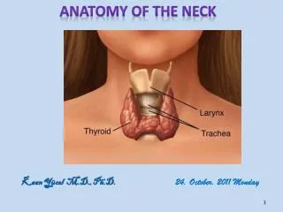
Thyroid Gland: Body's largest endocrine gland Is deep to sternothyroid and sternohyoid M At C5-T1 level. It has a capsule and externally covered by a sheath. Consists of an isthmus (1), which unites the lobes. A right lobe (2) and a left lobe (3). Pyramidal lobe: A remnant of thyroglossal duct may persist in the middle part of thyroid (50%). Ectopic Thyroid ****Function: Produces Thyroxin which controls the rate of metabolism of the body, and Calcitonin controlling Ca++ metabolism. Blood supply: Superior (8) and inferior (9) thyroidal artery
Goiter: Enlargement of Thyroid gland (nonneoplastic and noninflammatory). Usually not upward shift. Endemic in areas deficient in Iodine in food. Swelling in the neck which may disturb trachea, esophagus and/or laryngeal nerves. Exophthalmic goiter is due to excessive production of thyroxin. Thyroidectomy: Removal of thyroid due to cancer. Subtotal due to preservation of Parathyroid glands and recurrent as well as superior laryngeal nerves. Inadvertent removal of parathyroid glands lead to tetany, severe convulsion and muscle spasm due to decrease in serum Ca++ and may lead to immediate respiratory failure.
Parathyroid glands: Usually 4, one upper and one lower gland per each thyroid lobe. They are external to thyroid capsule and internal to the connective tissue sheath. Function: Produce parahormon, controlling the metabolism of P and Ca++.