Download
1 / 35
350 likes | 385 Views
Dive into the epidemiology, spectrum, and differential diagnosis of infected diabetic foot ulcers, challenges in diagnosing osteomyelitis, and effective evaluation strategies. Understand the importance of proper diagnosis, the golden standards such as bone biopsy, and cautious interpretation of diagnostic tests. Explore optimal treatment options, including antibiotics and the comparison between IV and oral therapies.

E N D
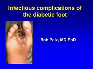
Infectious complications of the diabetic foot Bob Pelz, MD PhD
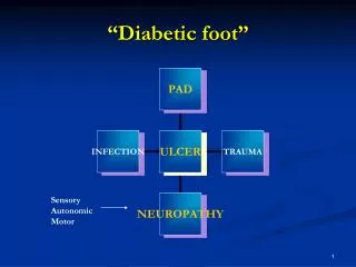
Epidemiology • 15% of diabetics develop ulcers, 6% require hospitalizaitons • Over half of ulcers become infected • 20-66% of infected ulcers involve bone
Spectrum of infections • Cellulitis • Abscess • Osteomyelitis
Differential diagnosis • Non-infected neuropathic ulcer • Fracture • Ischemia • Embolization, vasculitis, stasis ulcer, carcinoma
Pathogenesis • Sensory neuropathy • Trauma, deformity • Autonomic neuropathy • Diminished sweat, dry, cracked skin • Hyperglycemia • Decreased neutrophil function • Arterial disease
Challenges in Diagnosis of Osteomyelitis • Neuropathic changes may resemble infection on MRI, other images • Superficial cultures correlate poorly with deep organisms, and may not reflect deep infection at all • Radiographic signs absent early • Bone biopsy invasive, expensive, inaccurate
Diagnosis of Osteomyelitis • Labs: ESR > 70 • Radiology • MRI, Labeled wbc, plain film • Probe to Bone • Bone biopsy for histopathology, Cx • Surface cultures • Wound > 2 cm2
Plain radiographs • Cheap and often very helpful • Moth-eaten necrotic bone is dead and requires surgery
Probe-to-bone • Grayson, JAMA 1995. 75 inpatients, 66% with osteomyelitis • “On gentle probing, the evaluator detected a rock-hard, often gritty structure without the apparent presence of any intervening soft tissue” • Gold standard- histo or clinical + radiology • Sens/spec/PPV/NPV: 66,85,89,56%
Probe to Bone • Lavery et al (Diab. Care 2007): 247 outpts, 12% with OM. • S / S / PPV / NPV=87 / 91 / 57 / 98. • Shone, et al Diab Care 2006 • Sensitivity / Specificity 0.38 / 0.91 • Aragon-Sanchez, Diab Med 2011 PTB or X ray +. Gold standard = Bx with path showing osteo • Sens / Spec 0.97 / 0.92. LR +/- 12.8 / 0.02 • 85% of those with pos path had pos Cx • With exposed bone or positive probe to bone, IDSA guidelines (2004) say X- ray not needed
Bone Bx • Gold standard in most studies • Open Bx more accurate than needle • 31 pts, both needle and open (Seneville, CID 2009) • 23.9% correlation between open Bx and needle Biopsy Cx • Highest with Staph aureus (46.7%) • 41.7 correlation between swab Cx and biopsy culture • 82.3 for Staph aureus
Bone Biopsy • Weiner (J Foot Ankle Surg 2011) 44 pts with clinical osteo. • Just as likely for Bx to be pos by micro as by histo • Pos Cx rate low- 34% of 41 histologic osteomyelitis • 4 pos Cx in 34 histo-neg pts (Wu et al AJR 2007) • White, et al (Radiology 1995) Culture swab sensitivity 42%. 50% of histo-positive Bx had positive Cx • Should send Bx specimens for both Cx and histo
Superficial cultures, pitfalls • Poorly predictive of deep pathogens • 44% of sinus tract Cx contained organism from surg sample (Mackowiak JAMA 1978) • 28% concordance, 38% for staph (Zuluaga BMC Infect Dis 2002) • Twice as many bacteria species isolated by swab than by Bx (Kessler, Diab Med 2005)
Superficial Cx, advantages • Can often choose ABX to cover all plausible organisms • Organisms isolated repeatedly and in large numbers likely to be causative • Useful for detecting MRSA, other MDRO • Staph aureus likely pathogen if found
Osteomyelitis diagnosis, Meta-analysis Butalia, et al. JAMA 2008
Osteomyelitis diagnosis, Meta-analysis Dinh, MT, CID 2008
Osteomyelitis Treatment • Aerobic GPCs are the predominant pathogens in diabetic foot infections • Broad-spectrum empirical therapy is not routinely required but is indicated for severe infections • Acute infections are often monomicrobial (almost always with aerobic GPC) Lipsky et al, CID, 2004
Microbiology Lipsky, et al. CID 2004
Antibiotics • Surgery vs abx vs both. • ABX can’t sterilize dead bone • IV vs po • Easier to monitor therapy with IV, especially through RIC or in SNF • IV may be preferable if litigious or unreliable pt • IV expensive, PICC risks (DVT, infection, etc.)
IV vs PO therapy • IV Cloxacillin vs Bactrim/rif, 50 pts with surgical Cx, RCT. (Euba AAC 2009) • Relapses no different with 7-9 years f/u • Gentry, et al (AAC 1991) Ofloxacin vs IV, Bx-confirmed osteo. • 74% vs 86% w/out relapse at 18 month f/u • Fleroxacin/rif vs IV: 89% vs. 69% cure (Schrenzel, CID 2004) • Ofloxacin/Rif: Diabetic foot Staph. osteo. 76% relapse free at 22 mo. (Senneville CID 2001)
IV vs PO therapy • 9/11 osteo cured with Rif/Linezolid vs 9/10 with Rif/Bactrim (Nguyen Clin Micro Infect 2009). Similar cure with infected hardware. • Linezolid vs Unasyn or vanco (MRSA). 45 sites, 8 countries. (Lipsky, CID 2004) Excluded ischemic feet. 371 pts. Cured osteo in 27/44 Linezolid, 11/16 unasyn. More AEs in L arm, but mild
IV vs PO therapy • Generally, cure rates with IV and po therapy comparable. Rifampin almost always given.
Duration of therapy • 4-6 weeks typical, but not based on randomized data • IV followed by 3 months po if inadequate debridement
Case • 60, dm, h/o right 4th and 5th ray amputations, retinopathy, neuropathy • 4/27/11- Fever, Acute red, tender foot. • MRI cuboid edema, ?5th met osteo. No abscess • Cx- Group B Strep • Keflex 1 week • Offloading
Case • 5/10/11 Foot red, 1 week off keflex • X ray- no osteo • CRP- 0.7 • 5/24 pus, CRP=9.7, Cx=GBS, faxed in 20 days doxycycline • 5/31 erythema better • 7/11 Total contact cast
Case, cont. • 8/1/11 Copious drainage, necrotic base, +/- PTB despite total contact cast • X ray- still no osteo. • Tagged WBC c/w osteo • TcPO2 42
Case, cont • To OR, 8/11/11 • Path- no osteo, but possible fracture • Cx- Proteus, enterococcus • 2 weeks keflex • Wound improving with resection of weight-bearing 5th metatarsal • Wound healed as of 9/11
Case summary • 8 ID, 3 ortho, 13 wound care encounters over 5 months • 3 X rays, 1 MRI, 1 bone/WBC scan, TCC, surg • Cellulitis, possible abscess, but osteo never definite clinically, probably never had it despite positive cultures. • Fracture vs infection • Ulcer due to abnormal weight bearing, resolved with surgery • Lives with son who is nearly blind
Take-homes • Diagnosis and management of infected foot ulcers difficult, requires team approach • Anaerobes, resistant gram negatives not as common as taught. Staph aureus is at least half of infections. • Swab Cx, probe to bone, X rays useful • Oral therapy likely as good as IV