Download

1 / 51
520 likes | 761 Views
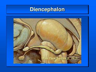
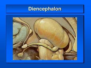
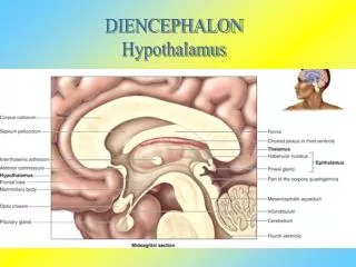
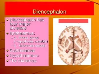
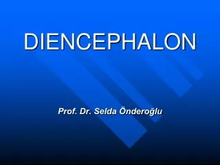
Diencephalon. Three paired structures Thalamus Hypothalamus Epithalamus Encloses the third ventricle. Cerebral hemisphere. Septum pellucidum. Corpus callosum. Interthalamic adhesion (intermediate mass of thalamus). Fornix. Choroid plexus. Thalamus (encloses third ventricle).

E N D
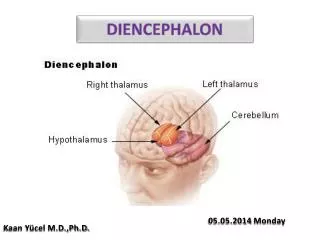
Diencephalon Three paired structures Thalamus Hypothalamus Epithalamus Encloses the third ventricle
Cerebral hemisphere Septum pellucidum Corpus callosum Interthalamic adhesion (intermediate mass of thalamus) Fornix Choroid plexus Thalamus (encloses third ventricle) Interven- tricular foramen Posterior commissure Pineal gland (part of epithalamus) Anterior commissure Corpora quadrigemina Mid- brain Cerebral aqueduct Hypothalamus Optic chiasma Arbor vitae (of cerebellum) Pituitary gland Fourth ventricle Mammillary body Choroid plexus Pons Cerebellum Medulla oblongata Spinal cord Figure 12.12
Thalamus 80% of diencephalon Superolateral walls of the third ventricle Connected by the interthalamic adhesion (intermediate mass) Contains several nuclei, named for their location Nuclei project and receive fibers from the cerebral cortex
Thalamic Function Gateway to the cerebral cortex Sorts, edits, and relays information Afferent impulses from all senses and all parts of the body Impulses from the hypothalamus for regulation of emotion and visceral function Impulses from the cerebellum and basal nuclei to help direct the motor cortices Mediates sensation, motor activities, cortical arousal, learning, and memory
Hypothalamus Forms the inferolateral walls of the third ventricle Contains many nuclei Example: mammillary bodies Paired anterior nuclei Olfactory relay stations Infundibulum—stalk that connects to the pituitary gland
Hypothalamic Function Autonomic control center for many visceral functions (e.g., blood pressure, rate and force of heartbeat, digestive tract motility) Center for emotional response: Involved in perception of pleasure, fear, and rage and in biological rhythms and drives
Hypothalamic Function Regulates body temperature, food intake, water balance, and thirst Regulates sleep and the sleep cycle Controls release of hormones by the anterior pituitary Produces posterior pituitary hormones
Epithalamus Most dorsal portion of the diencephalon; forms roof of the third ventricle Pineal gland—extends from the posterior border and secretes melatonin Melatonin—helps regulate sleep-wake cycles
Cerebral hemisphere Septum pellucidum Corpus callosum Interthalamic adhesion (intermediate mass of thalamus) Fornix Choroid plexus Thalamus (encloses third ventricle) Interven- tricular foramen Posterior commissure Pineal gland (part of epithalamus) Anterior commissure Corpora quadrigemina Mid- brain Cerebral aqueduct Hypothalamus Optic chiasma Arbor vitae (of cerebellum) Pituitary gland Fourth ventricle Mammillary body Choroid plexus Pons Cerebellum Medulla oblongata Spinal cord Figure 12.12
Brain Stem Three regions Midbrain Pons Medulla oblongata
Brain Stem Similar structure to spinal cord but contains embedded nuclei Controls automatic behaviors necessary for survival Contains fiber tracts connecting higher and lower neural centers Associated with 10 of the 12 pairs of cranial nerves
Frontal lobe Olfactory bulb (synapse point of cranial nerve I) Optic chiasma Optic nerve (II) Optic tract Mammillary body Midbrain Pons Temporal lobe Medulla oblongata Cerebellum Spinal cord Figure 12.14
View (a) Optic chiasma Optic nerve (II) Diencephalon Crus cerebri of cerebral peduncles (midbrain) • Thalamus • Hypothalamus Thalamus Diencephalon Mammillary body Hypothalamus Midbrain Oculomotor nerve (III) Pons Brainstem Trochlear nerve (IV) Medulla oblongata Trigeminal nerve (V) Pons Middle cerebellar peduncle Facial nerve (VII) Abducens nerve (VI) Vestibulocochlear nerve (VIII) Glossopharyngeal nerve (IX) Hypoglossal nerve (XII) Pyramid Vagus nerve (X) Ventral root of first cervical nerve Accessory nerve (XI) Decussation of pyramids Spinal cord (a) Ventral view Figure 12.15a
Crus cerebri of cerebral peduncles (midbrain) Thalamus View (b) Infundibulum Superior colliculus Pituitary gland Inferior colliculus Trochlear nerve (IV) Superior cerebellar peduncle Trigeminal nerve (V) Pons Middle cerebellar peduncle Facial nerve (VII) Inferior cerebellar peduncle Abducens nerve (VI) Vestibulocochlear nerve (VIII) Glossopharyngeal nerve (IX) Olive Hypoglossal nerve (XII) Thalamus Vagus nerve (X) Diencephalon Hypothalamus Midbrain Accessory nerve (XI) Pons Brainstem Medulla oblongata (b) Left lateral view Figure 12.15b
Thalamus View (c) Diencephalon Midbrain • Superiorcolliculus Corpora quadrigemina of tectum • Inferiorcolliculus • Trochlear nerve (IV) Pineal gland • Superior cerebellar peduncle Pons • Middle cerebellar peduncle Medulla oblongata Anterior wall of fourth ventricle • Inferior cerebellar peduncle • Facial nerve (VII) • Vestibulocochlear nerve (VIII) • Glossopharyngeal nerve (IX) Choroid plexus (fourth ventricle) • Vagus nerve (X) • Accessory nerve (XI) Dorsal median sulcus Thalamus Dorsal root of first cervical nerve Diencephalon Hypothalamus Midbrain Pons Brainstem (c) Dorsal view Medulla oblongata Figure 12.15c
Midbrain Located between the diencephalon and the pons Cerebral peduncles Contain pyramidal motor tracts Cerebral aqueduct Channel between third and fourth ventricles
Pons Forms part of the anterior wall of the fourth ventricle Fibers of the pons Connect higher brain centers and the spinal cord Relay impulses between the motor cortex and the cerebellum Origin of cranial nerves V (trigeminal), VI (abducens), and VII (facial) Some nuclei of the reticular formation Nuclei that help maintain normal rhythm of breathing
Medulla Oblongata Joins spinal cord at foramen magnum Forms part of the ventral wall of the fourth ventricle Contains a choroid plexus of the fourth ventricle Pyramids—two ventral longitudinal ridges formed by pyramidal tracts Decussation of the pyramids—crossover of the corticospinal tracts
Medulla Oblongata Respiratory centers Generate respiratory rhythm Control rate and depth of breathing, with pontine centers
Medulla Oblongata Additional centers regulate Vomiting Hiccuping Swallowing Coughing Sneezing
The Cerebellum 11% of brain mass Dorsal to the pons and medulla Subconsciously provides precise timing and appropriate patterns of skeletal muscle contraction
Anatomy of the Cerebellum Two hemispheres connected by vermis Each hemisphere has three lobes Anterior, posterior, and flocculonodular Folia—transversely oriented gyri Arbor vitae—distinctive treelike pattern of the cerebellar white matter
Anterior lobe Cerebellar cortex Arbor vitae Cerebellar peduncles Posterior lobe • Superior • Middle Choroid plexus of fourth ventricle • Inferior Medulla oblongata Flocculonodular lobe (b) Figure 12.17b
Anterior lobe Posterior lobe Vermis (d) (d) Figure 12.17d
Cerebellar Peduncles All fibers in the cerebellum are ipsilateral Three paired fiber tracts connect the cerebellum to the brain stem Superior peduncles connect the cerebellum to the midbrain Middle peduncles connect the pons to the cerebellum Inferior peduncles connect the medulla to the cerebellum
Cerebellar Processing for Motor Activity Cerebellum receives impulses from the cerebral cortex of the intent to initiate voluntary muscle contraction Signals from proprioceptors and visual and equilibrium pathways continuously “inform” the cerebellum of the body’s position and momentum Cerebellar cortex calculates the best way to smoothly coordinate a muscle contraction A “blueprint” of coordinated movement is sent to the cerebral motor cortex and to brain stem nuclei
Cognitive Function of the Cerebellum Recognizes and predicts sequences of events during complex movements Plays a role in nonmotor functions such as word association and puzzle solving
Functional Brain Systems Networks of neurons that work together and span wide areas of the brain Limbic system Reticular formation
Limbic System Structures on the medial aspects of cerebral hemispheres and diencephalon Includes parts of the diencephalon and some cerebral structures that encircle the brain stem
Fiber tracts connecting limbic system structures Septum pellucidum Diencephalic structures of the limbic system Corpus callosum •Fornix •Anterior thalamic nuclei (flanking 3rd ventricle) •Anterior commissure Cerebral struc- tures of the limbic system •Hypothalamus •Mammillary body •Cingulate gyrus •Septal nuclei •Amygdala •Hippocampus •Dentate gyrus •Parahippocampal gyrus Olfactory bulb Figure 12.18
Limbic System Emotional or affective brain Amygdala—recognizes angry or fearful facial expressions, assesses danger, and elicits the fear response Cingulate gyrus—plays a role in expressing emotions via gestures, and resolves mental conflict Puts emotional responses to odors Example: skunks smell bad
Limbic System: Emotion and Cognition The limbic system interacts with the prefrontal lobes, therefore: We can react emotionally to things we consciously understand to be happening We are consciously aware of emotional richness in our lives Hippocampus and amygdala—play a role in memory
Reticular Formation Three broad columns along the length of the brain stem Raphe nuclei Medial (large cell) group of nuclei Lateral (small cell) group of nuclei Has far-flung axonal connections with hypothalamus, thalamus, cerebral cortex, cerebellum, and spinal cord
Reticular Formation: RAS and Motor Function RAS (reticular activating system) Sends impulses to the cerebral cortex to keep it conscious and alert Filters out repetitive and weak stimuli (~99% of all stimuli!) Severe injury results in permanent unconsciousness (coma)
Reticular Formation: RAS and Motor Function Motor function Helps control coarse limb movements Reticular autonomic centers regulate visceral motor functions Vasomotor Cardiac Respiratory centers
Radiations to cerebral cortex Visual impulses Auditory impulses Reticular formation Descending motor projections to spinal cord Ascending general sensory tracts (touch, pain, temperature) Figure 12.19
Brain Waves Patterns of neuronal electrical activity Generated by synaptic activity in the cortex Each person’s brain waves are unique Can be grouped into four classes based on frequency measured as Hertz (Hz)
Types of Brain Waves Alpha waves (8–13 Hz)—regular and rhythmic, low-amplitude, synchronous waves indicating an “idling” brain Beta waves (14–30 Hz)—rhythmic, less regular waves occurring when mentally alert Theta waves (4–7 Hz)—more irregular; common in children and uncommon in adults Delta waves (4 Hz or less)—high-amplitude waves seen in deep sleep and when reticular activating system is damped, or during anesthesia; may indicate brain damage
Consciousness Conscious perception of sensation Voluntary initiation and control of movement Capabilities associated with higher mental processing (memory, logic, judgment, etc.) Loss of consciousness (e.g., fainting or syncopy) is a signal that brain function is impaired
Consciousness Clinically defined on a continuum that grades behavior in response to stimuli Alertness Drowsiness (lethargy) Stupor Coma
Sleep State of partial unconsciousness from which a person can be aroused by stimulation Two major types of sleep (defined by EEG patterns) Nonrapid eye movement (NREM) Rapid eye movement (REM)
Sleep First two stages of NREM occur during the first 30–45 minutes of sleep Fourth stage is achieved in about 90 minutes, and then REM sleep begins abruptly
Importance of Sleep Slow-wave sleep (NREM stages 3 and 4) is presumed to be the restorative stage People deprived of REM sleep become moody and depressed REM sleep may be a reverse learning process where superfluous information is purged from the brain Daily sleep requirements decline with age Stage 4 sleep declines steadily and may disappear after age 60
Language Language implementation system Basal nuclei Broca’s area and Wernicke’s area (in the association cortex on the left side) Analyzes incoming word sounds Produces outgoing word sounds and grammatical structures Corresponding areas on the right side are involved with nonverbal language components
Memory Storage and retrieval of information Two stages of storage Short-term memory (STM, or working memory)—temporary holding of information; limited to seven or eight pieces of information Long-term memory (LTM) has limitless capacity
Outside stimuli General and special sensory receptors Afferent inputs Temporary storage (buffer) in cerebral cortex Data permanently lost Data selected for transfer Automatic memory Forget Short-term memory (STM) Forget Data transfer influenced by: Excitement Rehearsal Association of old and new data Retrieval Long-term memory (LTM) Data unretrievable Figure 12.22
Transfer from STM to LTM Factors that affect transfer from STM to LTM Emotional state—best if alert, motivated, surprised, and aroused Rehearsal—repetition and practice Association—tying new information with old memories Automatic memory—subconscious information stored in LTM
Categories of Memory Declarative memory (factual knowledge) Explicit information Related to our conscious thoughts and our language ability Stored in LTM with context in which it was learned
Categories of Memory Nondeclarative memory Less conscious or unconscious Acquired through experience and repetition Best remembered by doing; hard to unlearn Includes procedural (skills) memory, motor memory, and emotional memory