Sheep Brain Dissection Guide
Sheep Brain Dissection Guide. Good Luck!!. Laurie Hayes. To begin. Get gloves and a rubber apron or lab coat if you would like. Goggles are also available for those who would like them. Pick up dissection tray, dull probe, pins, scissors, and scalpel

Sheep Brain Dissection Guide
E N D
Presentation Transcript
Sheep Brain DissectionGuide Good Luck!! Laurie Hayes
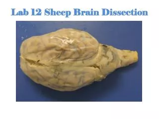
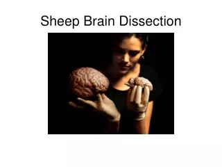
To begin • Get gloves and a rubber apron or lab coat if you would like. Goggles are also available for those who would like them. • Pick up dissection tray, dull probe, pins, scissors, and scalpel • Obtain a sheep brain and share this with a partner (or 2) • Rinse the sheep brain to remove preservative
Meninges of the Brain The brain is protected by the skull and 3 layers of membranes called meninges
Observe Meninges • Examine the brain to see if there is a tough outer coat…This is the DURA MATER (literally means “tough mother”) • Now look for the stringy, web-like ARACHNOID MATER (literally means spider mother) beneath and stuck to the dura mater. The space under the arachnoid, the subarachnoid space, is filled with cerebrospinal fluid and contains blood vessels. • Look deeper and see an thin transparent membrane that follows the contour of the ridges (gyri) and valleys (sulci) of the brain. This is the PIA MATER (means tender mother) • *remove the meninges
Questions?? 1. What is the function of the meninges? 2. Describe the following meninges: Dura mater, Arachnoid mater, and Pia mater • What is meningitis?
Main Brain Regions • Identify the cerebrum, brain stem and cerebellum on your brain 4. Draw picture of sheep brain and label structures Cerebrum Cerebellum Brain stem
Questions?? 5. Which of the previous parts of the brain do the following? a) Balance b) Breathing c) Hearing and Vision
Human vs Sheep • Compare the various areas of the sheep brain (cerebrum, brain stem, cerebellum) to the human brain. 6. How is it the same? 7. How is it different? Human brain
Identify Dorsal Structures • With the dorsal side up, identify the cerebral hemispheres, gyri, sulci,and longitudinal fissure on your sheep brain
Questions?? • What does the longitudinal fissure divide? • What is the difference between gyri and sulci? • What is the purpose of the gyri and sulci?
Lobes of the Cerebrum • Find the 4 lobes of the brain-frontal, parietal, occipital, and temporal • Using pins label each lobe • Teacher check & get my initials
Questions???? • Name the cerebral lobe that functions in: a) Hearing b) Vision c) Touch d) Movement
Corpus Callosum • At the longitudinal fissure, gently separate the two hemispheres and look down between them for the thick band of white fibers. This is the corpus callosum. • What is its function?
Identify Ventral Structures • Place the sheep brain ventral side up. • Identify the olfactory bulb, optic nerve, optic chiasm • Using pins label each structure. • Teacher check & initials 15. Draw and label the structures on your paper
Questions?? • How does the human brain’s olfactory bulb differ from the sheep’s? • Which is able to smell more odors? • Look at the optic chiasma. What happens to the optic nerve (vision) at the optic chiasma? • Which side of the occipital lobe (right or left) “sees” for the right eye?
Brain Stem • Look at the three parts of the brain stem-the midbrain (cerebral peduncle and mammillary body), pons and medulla oblongata • Pin and label parts • Teacher check & initials
Function • Mammillary bodies serve as relay stations for reflexes related to sense of SMELL • Cerebral Peduncle-group of myelinated nerves
Questions?? • What is the function of the pons? • What is the function of the medulla oblongata? • Could you survive without them? • Why or Why not?
Identify dorsal structures • Carefully pull the cerebellum away from the cerebrum. Don’t break any structures off. • Identify the pineal body, superior colliculi, and inferior colliculi.
Functions-Reflex Centers • Inferior Colliculi- movement of head and trunk in response to sound stimuli • Superior Colliculi-movement of eyes, head and neck in response to visual stimuli
Pineal Body (Gland) • Produces the hormone melatonin at night • Melatonin is the sleep hormone
Questions?? • What is the function of the Pineal body?
Midsagittal Section • Cut your sheep brain in half • Locate the brain stem parts: the medulla oblongata, the pons, cerebrum, and cerebellum
Cerebellum • Look at the cross section of the cerebellum. The inner structure is called the arbor vitae (living tree). 26. Why is this a good name for it?
Midsagittal Section • Locate the corpus callosum, thalamus,lateral ventricle, fourth ventricle and cerebral aqueduct
Questions 27. What is the function of the ventricles? 28. The cerebral aquaduct allows for the circulation of the cerebral spinal fluid through the ventricles. What do you think would happen if the duct became blocked? 29. How would that affect brain tissue?
The End!! Good bye or in sheep language BAA BAA