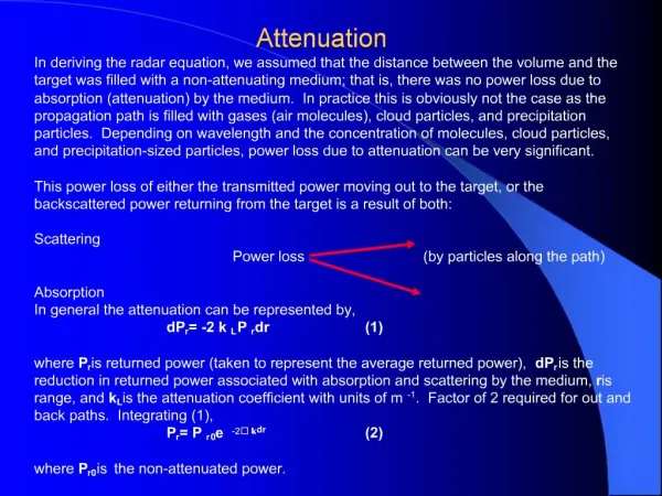
Attenuation
Attenuation. Attenuation. Weakening of the wave due to interaction with the medium in intensity & amplitude of the sound wave Measured in decibels (-dB; negative sign is assumed when referring to attenuation). Attenuation.


Attenuation
E N D
Presentation Transcript
Attenuation • Weakening of the wave due to interaction with the medium • in intensity & amplitude of the sound wave • Measured in decibels (-dB; negative sign is assumed when referring to attenuation)
Attenuation • Affects the image (must be compensated for through the components & controls of the US unit) • Without understanding attenuation effects, it is not possible to understand how & why the US system controls work
3 Types of Attenuation • Absorption • Reflection • Refraction
Absorption • conversion of sound energy into heat within the medium
CRITICAL POINTS ABOUT ABSORPTION • Main form of attenuation in soft tissue • Accounts for about 80% of a sound beam’s attenuation in soft tissue • the amount of energy to reflect an echo; This reduces penetration & signal strength • Absorbed heat energy causes tissue heating, a potential risk of tissue damage exponentially with frequency
3 Types of Attenuation • Absorption • Reflection • Refraction
Anatomy of Reflection • When sound waves meet an interface between 2 media (i.e. blood % vessel wall) • Part is reflected back to the transducer ( the rest of the waves’ intensity) • Remaining waves propagate through the interface into the next medium • There are 3 parts of the sound beam when it encounters an interface
Incident Beam The incident beam is the red arrow & all its intensity that you start out with It strikes the interface between 2 different media
Reflected Beam The reflected beam is the open red arrow is the part of the sound beam coming back (echoing) towards the probe with its reflected intensity after it struck the interface between two different media
Transmitted Beam The remaining part of the beam that continues on traveling in the same direction after striking the boundary between two media is called the transmitted beam (goofy green arrow)
The type of reflection that occurs at tissue boundaries impacts… • Amount of energy reflected back to the probe • The return signal’s strength depends on how much of the wave energy is reflected back & received by the transducer: the stronger the backscatter, the stronger the signal • If too much reflection occurs at one boundary, there will not be enough energy left to visualize deeper tissue boundaries
Think that over a bit . . . (click here) Could this be the reason why you may select a different frequency transducer? Could this also be why we have difficulty seeing past air-filled structures like lung and bowel? Is this why calcium deposits and bone shadow???
Types of Reflection • Absorption • Reflection • Specular Reflection • (Back) Scattering • Rayleigh Scattering • Refraction
Angle of Reflection • Angle of the incident sound wave (beam) is measured from an imaginary line perpendicular to the interface (0°) • Angle of reflection occurs on the other side of the line perpendicular to the interface and is also measured from 0° • Angle of incidence = angle of reflection
Incidence Angle Reflection Angle 0° - Normal
Perpendicular (Normal) Incidence • Incident sound wave (beam) strikes the interface at a 0° angle (direction of wave is perpendicular to the interface) • Angle of reflection is the same angle the wave entered at (0°) • Angle of transmission is the same angle the wave entered at (0°)
Specular Reflection • Reflection that occurs from the sound wave striking a surface that is large, flat & smooth compared to the wavelength • Angle of incidence = angle of reflection
Types of Reflection • Absorption • Reflection • Specular Reflection • (Back) Scattering • Rayleigh Scattering • Refraction
(Back) Scattering Wave reflected back at many different directions. This type of reflection occurs from surfaces that are rough in respect to the wavelength.
Types of Reflection • Absorption • Reflection • Specular Reflection • (Back) Scattering • Rayleigh Scattering • Refraction
Rayleigh Scattering • Occurs from structures that are small relative to the wavelength • This is frequency-dependent scattering Recall: Wavelength changes with changes in frequency. Changing the frequency may make the wavelength smaller or greater in size compared to the structure that always remain the same size.
Facts about Reflection • Body surfaces are “rough” compared to the • produce scattering • provides the tissue ‘texture’ • Bones, diaphragm & gas are strong specular reflectors cause imaging artifacts • Size of red blood cells (RBC’s) < λ ;a Rayleigh scatterer
Facts about Reflection • Reflections from a non-perpendicular angle of incidence may not ever bounce back to the transducer • Changes in the angle can affect the display intensity & produce artifacts • Images are best when the angle of incidence is perpendicular (normal) to the structure’s surface
Examples of Typical US Reflection • 1% of the beam - reflected at a soft tissue/soft tissue interface • 30-50% of the beam - reflected at a soft tissue/bone interface • 99% of the beam - reflected at a soft tissue/air interface
What would the reflected dB be if the initial beam intensity is 50 dB? The intensity of the reflected beam from ST is only 1% of the original intensity. 30 dB RECALL: 1% = 1/100 In dB that = -20dB (learned in Session 2!) 50 dB -20dB = 30 dB
What would the reflected dB be if the initial beam intensity is 50 dB? 30-50% of the beam is reflected at a soft tissue/bone interface – so let’s assume 40% as an average 34dB RECALL: 40% = ¼ X 1/10 In dB that = -6dB + -10 dB or -16dB 50 dB -16dB = 34 dB
Notice the dB range between the 2 answers doesn’t seem that great.Actually it is because dB is a way making a large range of numbers into a smaller easier way to manage numbers. • If you are really daring, try to calculate the problem using bone (hint – it is really close to 50dB, which is why we don’t see behind them very well, most of the beam reflects)
Acoustic Impedance (Z) • Resistance to sound travel in a medium (Z) • Caused by a mismatch between the 2 mediums’ propagation speeds • Amount of reflection depends on how mismatched the mediums’ density & speed are • No Z mismatch - “0” reflection & 100% transmission • Large Z mismatch - large amt. of reflection & small amt. of transmission • Unit is Rayls (Z)
Acoustic Impedance (Z) Acoustic Impedance (Rayls) = (density) * (sound velocity) 1 (Rayl) = 1 (kg/m3) * 1 (m/sec) = kg / (m2 * sec) Z = p * c
Z values You want ever be asked to calculate a Z value (unless you become a physicist!) Just correlate how the acoustic impedance of one tissue relates to another tissue’s impedance value. In the following, Z1 is the impedance value of the first tissue and Z2 is the second tissue
Intensity Reflection Coefficient (IRC) Ireflected Z2 - Z1 Itotal Z2+ Z1 IRC% reflected = 2 Determines the fraction of the initial intensity that gets reflected difference between Z1 & Z2 = IRC
Intensity Transmission Coefficient (ITC) ITC% reflected = 2 Ireflected Z2 - Z1 Itotal Z2+ Z1 1 - Determines the fraction of the initial intensity that gets transmitted into the 2nd medium
Example What happens if: Z2 = Z1 (no impedance mismatch)? If there is no acoustic impedance mismatch there will be ‘0’ (zero) reflection or only 100% transmission.
What happens when sound goes from: Soft tissue to bone (4080 m/s)? Air (330 m/s) to soft tissue? When Z2 is significantly greater than Z1, the greater the amount of reflection
What happens when sound . . . goes from air to ST? How effective is scanning if you try to transmit US through a layer of air into tissue (like the skin without gel)? How do you think Zgel compares with Zair?
Answer Zair (.0004) < Ztransducer (29.0) Result - very large Z mismatch - very little transmission Zgel > Zair; this mismatch allows for more sound transmission into the body This is the real reason to use gel, it serves as an intermediate layer between 2 mismatching mediums.
Points on Attenuation = = attenuation ( absorption) = penetration (less energy transmitted) distance = attenuation
Determining Attenuation(Attenuation Coefficient) Dependent on frequency & the medium soft tissue: 0.5 dB / (cm-MHz) muscle: 1.0 dB / (cm-MHz) blood: 0.125 dB / (cm-MHz)
Examples of Attenuation Coefficient for soft tissue: 0.5 dB / (cm-MHz) • What is the attenuation of a 10MHz US beam at a depth of 10 cm in soft tissue? (0.5 dB) x (10 cm) x (10 MHz) = 50 dB cm-MHz • What is the attenuation of a 10MHz US beam imaging a depth of 10 cm in soft tissue? (0.5 dB) x (20 cm) x (10 MHz) = 100 dB cm-MHz
Explanation • Attenuation of the beam going through ST • Attenuation of the beam going through & then returning back to the transducer (round-trip). In order to image the structure, the transducer must receive the returning echoes, thus the sound travels 2X the distance. The beam intensity for imaging a 10 cm depth is 1/10,000,000,000 of the original beam intensity.
Half Value Layer Thickness -Depth when the intensity is ½ of its original value (AKA penetration depth, half boundary layer) Depth (cm) = 3 Attenuation Coefficient
3 Types of Attenuation • Absorption • Reflection • Refraction
Refraction We have discussed perpendicular or normal incidence earlier in this presentation. To understand refraction, we need to know that any incidence wave that is not normal is termed as oblique incidence.
Oblique Incidence • Incident sound wave strikes the interface at a non-0° angle (direction of wave is not perpendicular to the interface) • Angle of reflection is the same angle the wave entered but on the other side of 0° • Angle of transmission can continue in the same direction of the incident beam or refract
Refraction • a change in direction or bending of the transmitted part of the sound wave as it passes through one medium into another • 2 conditions; both must be met : 1) Different propagation speeds (c) of the 2 media 2) Oblique incidence between the sound wave & the interface
Snell’s Law • Determines the refraction angle of sound transmitted through 2 media propagation speed (medium 1) =Sine incident angle propagation speed (medium 2) Sine transmission angle ci sin (θi) ct sin (θt)