Download

1 / 23
230 likes | 342 Views
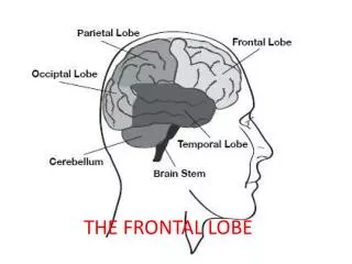
JHU BME 580.422 Biological Systems II Motor areas of the frontal lobe Reza Shadmehr. Summary of functions Posterior parietal cortex : align proprioception of arm with vision of hand compute hand position in fixation coordinates. compute target position in fixation coordinates.

E N D
JHU BME 580.422 Biological Systems II Motor areas of the frontal lobe Reza Shadmehr
Summary of functions • Posterior parietal cortex: • align proprioception of arm with vision of hand • compute hand position in fixation coordinates. • compute target position in fixation coordinates. • Premotor cortex: • Subtract target position with respect to hand position and code the desired movement in terms of displacement of the hand. • Primary motor cortex: • Transform the desired movement to muscle activity patterns and send this command to the spinal cord.
Arm motor commands Motor cortex Arm proprioception Difference vector (target location with respect to hand) Premotor cortex Fixation-centered location of hand Fixation-centered location of target Post. Parietal Cortex Arm position in proprioceptive coordinates Retinocentric location of target Eye and head orientation in proprioceptive coordinates
Coding object position with respect to the arm takes place in the premotor cortex A C B Graziano MSA et al. (1997) J Neurophysiol
In the premotor cortex, target of a reaching movement is coded with respect to the end-effector Kakei, Hoffman & Strick Science 1999 and Nature Neurosci 2001 Neural discharge rate Movement direction
Neurons in the primary motor cortex are sensitive to forces that are necessary to perform a reaching movement Kakei, Hoffman & Strick Science 1999 and Nature Neurosci 2001 Movement direction Neural discharge rate
Neuron 1 Neuron 2 Neuronal activity among some cells in the primary motor cortex relates to muscle forces Cabel, Cisek & Scott J Neurophysiol 2001 Experiment: A constant torque was applied to elbow and shoulder joints of the monkey’s arm. The animal is trained to maintain constant arm position. Therefore, muscles produce activity to counter the imposed torque. Neural activity is averaged over 2 seconds. Neural activity for many cells varies with direction of torque. Different cells have different preferred directions of torque.
During a reaching movement, activity in premotor cortex cells precedes the activity in M1 Moran & Schwartz, J Neurophys 1999
Stimulation of cortical surface in an individual Penfield & Rasmussen The Cerebral Cortex of Man (1950), Redrawn by M.H. Schieber J Neurophysiol 2001
Intracortical microstimulation maps of monkey motor cortex In anesthetized monkey, a microelectrode is positioned closed to layer V. Low current stimulation is delivered to excite ~30 pyramidal neurons. Flick-like twitches of discrete parts of the contralateral side of the body are observed. There is a general trend for somatotopy: trunk more medial, jaw more lateral. However, movement of a given body part (e.g., digits) is evoked from multiple foci. HC Kwan et al. J Neurophysiol 41:1120 1978, redrawn by Shieber J Neurophysiol 2001.
Damage to peripheral nerves causes change in the motor map In adult rat, motor map can change a few hours after a branch of the nerve supplying motor axons to the muscle attached to whiskers (vibrissae) is cut. No sensory fibers are damaged.
Neighboring regions of the motor cortex are connected via inhibitory interneurons. Damage to the peripheral nerve, reduces the synaptic efficacy of the inhibitory pathway from vibrissae to the forelimb. Stimulation in vibrissae area now causes motion of the forelimb. _ forelimb + + vibrissae
Amputation changes the motor map This individual’s right arm above the wrist has been amputated. The motor cortex is stimulated (via TMS) and muscle activity is recorded in biceps and depressor labii inferioris. The lip, and first and fifth digits are touched and evoked potentials are recorded from the brain. Contralateral to the stump Contralateral to the intact arm Motor representation of biceps contralateral to the stump is larger than on the side of the intact arm. Karl A. et al. (2001) J Neurosci 21:3609
Phantom Limb Pain (PLP) After amputation, the individual is likely to experience chronic pain in the missing limb. The pain is more common in the initial years after amputation, but may remain for many years. Patients with PLP tend to have an imbalance in the size of the motor map between the hemispheres. Amputated hand Right Right Left Biceps region Biceps region Left Right Left Karl A. et al. (2001) J Neurosci 21:3609
PLP may be related to mis-calibrated internal models • The brain relies on internal models to predict sensory consequences of movements. • Internal models adapt when there is a discrepancy between expected and actual sensory feedback. • In amputation, internal models must adapt in response to very large errors.
Sometimes the pain associated with the missing limb may be related to the fact that the patient’s motor system send commands to the missing limb to move it, but it does not receive feedback indicating that it has moved. To test this, one can place a mirror so that the patient views their normal limb and then ask the patient to make symmetric movements like clapping or conducting an orchestra while looking at the mirror. The patient may feel that the missing limb is finally moving, and in some cases this produces relief of the spasms and awkward posture that was causing the pain. Results: Greatest benefit to patients soon after amputation. Daily use significantly reduced the perception of pain. Control group Main group McCabe et al. (2003) Rheumatology 42:97-101
Effect of an infarct A lesion in the monkey motor cortex destroys 21% of digit and 7% of wrist. After 3 months, the area for digits has been reduced in neighboring regions as well. Before lesion 3 months post lesion Nudo & Milliken, J Neurophys 1996
Lesion destroyed 22% of digit region. After rehabilitation, the spared digit area increased by 15%. Nudo et al. Science 1996
Constrained motion rehabilitation The unaffected arm is restrained for 8 hours a day, 12 days. During this period, the affected arm is trained. Liepert, J. et al. Stroke 2000
Digit,wrist, and forearm Nudo et al. Sem Neurosci 1997
Summary of functions • Posterior parietal cortex: • align proprioception of arm with vision of hand • compute hand position in eye-centered coordinates. • compute target position in eye-centered coordinates. • Premotor cortex: • Subtract target position with respect to hand position and code the desired movement in terms of displacement of the hand. • Primary motor cortex: • Transform the desired movement to muscle activity patterns and send this command to the spinal cord.