Download
1 / 6
60 likes | 142 Views
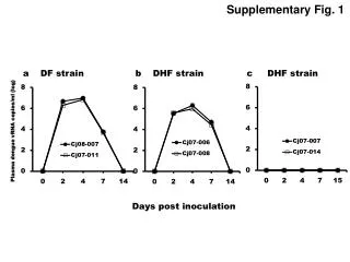
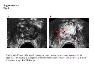
1B. 1A. 1D. 1C. Supplementary Fig. 1. Treatment of VEGF-C with the human acute myeloid leukemic cell line,THP-1,

E N D
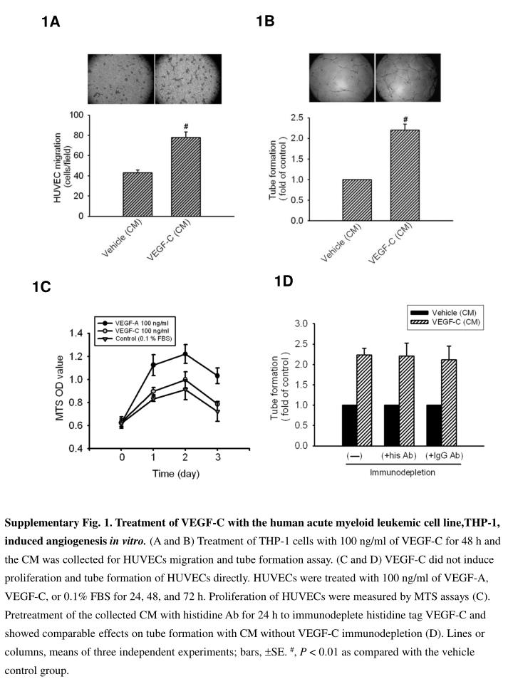
1B 1A 1D 1C Supplementary Fig. 1. Treatment of VEGF-C with the human acute myeloid leukemic cell line,THP-1, induced angiogenesis in vitro. (A and B) Treatment of THP-1 cells with 100 ng/ml of VEGF-C for 48 h and the CM was collected for HUVECs migration and tube formation assay. (C and D) VEGF-C did not induce proliferation and tube formation of HUVECs directly.HUVECs were treated with 100 ng/ml of VEGF-A, VEGF-C, or 0.1% FBS for 24, 48, and 72 h. Proliferation of HUVECs were measured by MTS assays (C). Pretreatment of the collected CM with histidine Ab for 24 h to immunodeplete histidine tag VEGF-C and showed comparable effects on tube formation with CM without VEGF-C immunodepletion (D).Lines or columns, means of three independent experiments; bars, SE. #, P < 0.01 as compared with the vehicle control group.
2B 2A Supplementary Fig. 2. Effects of COX-2 inhibitors, NS398 and indomethacin, on VEGF-C- induced angiogenesis in vitro. (A and B) THP-1 cells were pretreated with 10 mM NS398 or indomethacin for 30 min before incubation with 100 ng/ml of VEGF-C for 48 h, then the CM was collected for tube formation and HUVEC migration assay. Columns, means of three independent experiments; bars, SE. *, P < 0.05; **, P < 0.01 as compared with the VEGF-C treatment group.
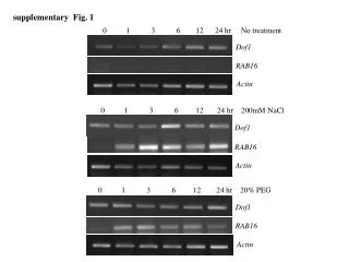
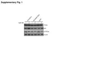
3A 3B Supplementary Fig. 3. VEGF-C and VEGF-A upregulate COX-2 expression in human acute myeloid leukemic cells. (A)COX-2 mRNA and protein expression were upregulated in a time-dependent fashion after VEGF-C (100 ng/ml) treatment, peaking at 8 and 48 h in 2 human acute myeloid leukemic cells, U937 and HL60. (B) COX-2 protein expression was upregulated in a time-dependent fashion after VEGF-A (50 ng/ml) treatment, peaking at 24 h in THP-1, U937, and HL60 cells.
Supplementary Fig. 4. Involvement of EP2 and EP4 receptors in the collected CM-induced tube formation. HUVECs tube formation ability of CM collected from VEGF-C-treated THP-1 after pretreatment of HUVECs with 3 mM AH6809 (EP2 antagonist), 30 mM AH23848 (EP4 antagonist), or 3 mM AH6809+30 mM AH23848 for 30 min. Columns, means of three independent experiments; bars, SE. **, P < 0.01 as compared with the VEGF-C treatment group.
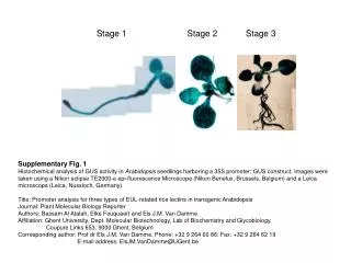
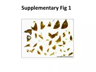
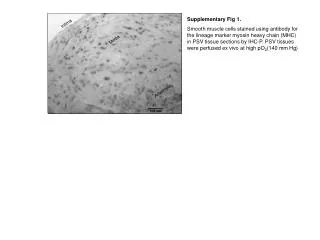
Supplementary Fig. 5. Similar localization of VEGF-C and COX-2. Immunohistochemical analysis of serial sections of bone marrow specimens from a representative patient with diagnosed AML shows identical coexpression and localization of VEGF-C and COX-2. Original magnification ×400.
Supplementary Table 1. Primers used to perform RT-PCR of the respective target genes