Download
1 / 37
380 likes | 679 Views
Documenting Heart Failure LR Brown, RN, MA, CCRN, CCDS, CDIP, CCS. Objectives. At the conclusion of this module, the learner will be able to: Differentiate between systolic and diastolic heart failure Describe three clinical indicators of acute heart failure Define acute cor pulmonale.

E N D
Documenting Heart FailureLR Brown, RN, MA, CCRN, CCDS, CDIP, CCS
Objectives • At the conclusion of this module, the learner will be able to: • Differentiate between systolic and diastolic heart failure • Describe three clinical indicators of acute heart failure • Define acute cor pulmonale
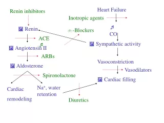
What is heart failure? • Heart, due to impairment of structure or function, does not pump effectively • Because oxygenated blood is not effectively pumped forward into the circulation, tissues do not receive nutrients and oxygen • Fluid backs up within the circulatory system leading to peripheral and pulmonary edema (“congestive heart failure”), • Most common cause is coronary artery disease
What’s the problem? • Documenting “CHF” is insufficient! • CMS guidelines require acuity and specificity. • Incomplete documentation of heart failure has a significant impact on severity of illness, risk of mortality, GLOS, and case mix index. • Incomplete documentation affects treatment modalities, core measures, and communication. • Incomplete documentation affects patient care.
But I wrote NYHA Class III… • Documenting NYHA classifications is not adequate documentation of heart failure for CMS
Is it systolic, diastolic, or combined? Specificity
Ejection fraction • EF = % of blood volume pumped out with each stroke • Usually refers to left ventricle, but right ventricle EF may also be measured • Commonly diagnosed by ECHOcardiogram, but may also be measured during stress test or cardiac catheterization • Ejection fraction is a key clue as to type of heart failure
Systolic heart failure • Ejection fraction < ~ 50% • Result of impaired inotropic state – heart can’t empty • Causes include dilated cardiomyopathy, myocardial infarction • Treated with ACE-inhibitors or ARBs – core measures; Digoxin, judicious use of β-blocker • Core measure HF-3: pts w/LV systolic dysfunction (EF < 40%) prescribed ARB or ACE-i
Diastolic heart failure • Ejection fraction > ~ 50 % • 1/3 of patients with symptomatic heart failure have “normal” EF • Risk increases with age • Result of impairment in heart’s ability to relax – heart can’t fill • May see elevated filling pressures “stiff ventricle” • Causes include restrictive and hypertrophic cardiomyopathy, ischemia, HTN, senile cardiac amyloid, constrictive pericarditis, myocardial ischemia • Treatment differs from systolic HF – β-blockade
Systolic and diastolic heart failure • It is possible to have elements of both systolic and diastolic heart failure • Physician should not write, “both,” or “combined,” because the coders can’t code from that • Physician needs to specify, “systolic and diastolic”
Acute heart failure • Usually – but not always – systolic • Develops suddenly without prior history • May have sudden reduction in cardiac output • May be hypotensive • May not show peripheral edema • Can be documented as “acute” or “decompensated” heart failure
Chronic heart failure • Develops slowly • May be seen in patients with dilated cardiomyopathy, valvular heart disease • Often has normal blood pressure but with peripheral edema • Patients coming from home with heart failure medications – if you continue those meds, are you treating a chronic condition?
Acute on chronic • Patient has pre-existing chronic condition that worsens • Can be documented either as “acute on chronic” or “exacerbation”
Look for clues to acuity and specificity • Check the BNP – is it elevated? • Check the ECHO results – what is the EF? How does the cardiologist describe LV function? • What are the pt’s clinical signs/symptoms? • Dyspnea, hypoxia • Rales/rhonchi • Peripheral edema • JVD • S3 gallop • How is the heart failure being treated? • IV diuresis usually means an acute condition! • No change in home HF meds usually means chronic
SI/IS for heart failure Condition-specific criteria
Heart failure admission guidelines • Need documentation of diagnosis, physical assessment to support diagnosis, documented evidence of cardiopulmonary instability, treatment per core measures (unless contraindicated), evaluation of LV systolic function, assessment of oxygenation and treatment with supplemental oxygen, use of cardiac monitoring • Heart failure can be monitored and treated in an observation status – not an automatic IP
Severity of illness & geometric length of stay Impact of documentation
Acuity/specificity vs “CHF” • “CHF” does nothing to increase relative weight and CMI • Acute heart failure, documented as systolic, diastolic, or systolic & diastolic, is a major co-morbidity that increases relative weight and CMI • Two patients with the same presentation and same treatment, documented differently, can have dramatically different relative weights, CMI, and geometric lengths of stay
Case 1 • Patient admitted with atrial fibrillation, develops signs of acute heart failure, EF is 60% • Physician documents AF, CHF • DRG 310, GLOS 2.0 days, RW 0.5608 • Physician documents AF, acute diastolic heart failure • DRG 308, GLOS 4.0 days, RW 1.2283
Case 2 • Patient admitted with intracranial hemorrhage, has been taking Lisinopril for previously diagnosed heart failure, meds are continued and patient placed on telemetry. ECHO done in June showed EF of 30% • Physician documents ICH, hx systolic dysfunction • DRG 66, GLOS 2.6 days, SOI 1, RW 0.8105 • Physician documents ICH, chronic systolic heart failure • DRG 65, GLOS 3.8 days, SOI 2, RW 1.8555
Case 3 • Patient admitted with CAD, hx systolic HF, undergoes CABG; postoperatively cannot handle fluid resuscitation, requires extensive diuresis and increased oxygen requirement, spends extra 2 days in the hospital • Physician documents CAD, fluid overload • DRG 236, GLOS 6.0 days, RW 3.7720 • Physician documents CAD, systolic HF exacerbation • DRG 235, GLOS 9.3 days, RW 5.9063
Getting acuity and specificity into the medical record Query process
Sample query: • Dr. Jones: “CHF” is documented in the medical record. The record indicates moderate dyspnea, bilateral rales and rhonchi. BNP is 9400. ECHO shows ejection fraction of 35%. Orders for oxygen, PO Lisinopril and IV Bumex. CMS requires acuity and specificity in documentation of heart failure. To establish the most accurate severity of illness of your patient, please specify the type (systolic, diastolic, or systolic & diastolic), and acuity (acute, chronic, or acute on chronic) of heart failure you are treating. For continuity of the record, please document in the progress notes and continue through to the discharge summary.
Corpulmonale • Type of right-sided heart failure • Acute cor pulmonale = acute lung disease (e.g., PE, ARDS, COPD exacerbation) causing acute right sided heart failure • Chronic cor pulmonale = right sided heart failure resulting from pulmonary hypertension, chronic lung disease, or pulmonary valve stenosis • Right ventricle dilates from chronic ischemia and hypertension of the arteries in the lungs • Ventricle unable to pump against the hypertension
Findings in corpulmonale • Acute onset of dyspnea • JVD • Hypotension • Hypoxia • Tachycardia • Shock • S3/S4 on inspiration • Fatigue • Exertional dyspnea • Ascites • Hepatomegaly • Dependent edema • Cardiomegaly • Syncope Acute Chronic
Diagnosing corpulmonale • CXR: RV and pulmonary artery enlargement • EKG: RV hypertrophy (R axis deviation, QR wave in V1, dominant R wave in V1 to V3) • Both CXR and EKG results may be skewed in COPD patient due to realignment of the heart • ECHO: evaluate LV and RV function, likely to see RV thickening and increased PA pressures • Often limited by lung disease • Right heart catheterization Cor Pulmonale, Merck Manual for Healthcare Professionals
Coding corpulmonale • Be aware of combination codes in ICD-10. • Not coded as heart failure. There is no code in heart failure DRGs for right sided heart failure. • As PDx, corpulmonale goes to DRG 316, other circulatory system diagnoses. • As SDx, acute corpulmonale is a major co-morbidity.
Pleural effusion • Can occur due to many causes • When found in patient with heart failure, is considered integral to disease and is not coded EXCEPT • Physician can state that the pleural effusion is clinically significant, apart from the heart failure, or is not due to the heart failure (look for evidence of different etiology for the pleural effusion) • If treatment of pleural effusion is not the same as that for heart failure (e.g., performing a thoracentesis—not routine tx for HF!), then the pleural effusion can also be coded
Review activity • Mrs. Gonzales is admitted with shortness of breath, rales in the bases; CXR indicates bilateral effusions, BNP is 12,400, EF is 28%; IV Lasix is ordered. The physician documents CHF with pleural effusion. Your most likely action is: • Code pleural effusion as PDx and query for acute systolic HF as SDx • Query for acute systolic HF as PDx and do not code the pleural effusion • Query for acute systolic HF as PDx and query for transudative pleural effusion as SDx • Code CHF as PDx and query whether the pleural effusion is related to the CHF
Review activity • On day 2, Mrs. Gonzales’s pulmonologist decides to perform a thoracentesis of the pleural effusion. Your most likely action is to: • Code the pleural effusion as PDx because that is the focus of the care • Not code the pleural effusion because it is inherent in heart failure • Code the pleural effusion as SDx because the treatment is outside the scope of heart failure • Query for acute respiratory failure due to the thoracentesis.
Review activity • A good way to remember the difference between systolic and diastolic heart failure is: • Systolic means the heart can’t fill; diastolic means the heart can’t pump • Systolic means the left ventricle is preserved; diastolic means the right ventricle is preserved • Systolic means the heart failure is acute; diastolic means the heart failure is chronic • Systolic means the heart can’t pump; diastolic means the heart can’t fill
Review activity • Acute cor pulmonale: • Is usually caused by an acute lung injury or disease • Is usually caused by acute systolic heart failure • Is best diagnosed by a left heart catheterization • Is never found in patients with chronic lung disease