Fig. 41-1
Fig. 41-1. Chapter 41 Animal Nutrition. Taken in. Taken apart. Taken up. Fig. 41-4. Fig. 41-6a. Fig. 41-6b. Leaf miner caterpillar, a substrate feeder. Humpback whale, a suspension feeder. Baleen. Rock python, a bulk feeder. Mosquito, a fluid feeder. Fig. 41-18. Evolutionary and
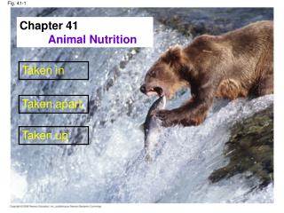

Fig. 41-1
E N D
Presentation Transcript
Fig. 41-1 Chapter 41 Animal Nutrition Taken in Taken apart Taken up
Fig. 41-6a Fig. 41-6b Leaf miner caterpillar,a substrate feeder Humpback whale, a suspension feeder Baleen Rock python, a bulk feeder Mosquito, a fluid feeder
Fig. 41-18 Evolutionary and Adaptations Incisors Molars Canines Premolars (a) Carnivore (b) Herbivore (c) Omnivore
Fig. 41-9 Crop Gizzard Variation in alimentary canals Intestine Esophagus Pharynx Anus Mouth Typhlosole Lumen of intestine (a) Earthworm Foregut Midgut Hindgut Esophagus Rectum Anus Crop Mouth Gastric cecae (b) Grasshopper Stomach Gizzard Intestine Mouth Esophagus Crop Anus (c) Bird
Stomach and intestinal adaptions Stomach Small intestine Small intestine Cecum Colon(largeintestine) Carnivore Herbivore Fig. 41-19
Mutualistic adaptation Fig. 41-20 蜂巢胃 Rumen Reticulum 1 2 瘤胃 Intestine Esophagus 重瓣胃 3 皺胃 Omasum Abomasum 4
Concept 41.2: The main stages of food processing are ingestion, digestion, absorption, and elimination Smallmolecules Piecesof food Chemical digestion(enzymatic hydrolysis) Mechanicaldigestion Nutrientmoleculesenter bodycells Undigestedmaterial Food Ingestion Digestion Elimination Absorption 2 4 1 3 • Ingestion is the act of eating
Fig. 41-10a Tongue Sphincter Oral cavity Salivary glands Pharynx Esophagus Sphincter Liver Stomach Ascendingportion oflarge intestine Gall-bladder Duodenum ofsmall intestine Pancreas Smallintestine Smallintestine Peristalsis sphincters Largeintestine Rectum Anus Appendix Cecum
Essential Nutrients • There are four classes of essential nutrients: • Essential amino acids • Essential fatty acids • Vitamins • Minerals
Fig. 41-2 Essential amino acids for adults Beans and otherlegumes Methionine Valine Threonine Phenylalanine Leucine Isoleucine Corn (maize)and other grains Tryptophan Lysine • insufficient essential amino acids----- • Malnutrition or called protein deficiency • Vegetarian diet---
Fig. 41-3 Storing protein for growth Muscle protein Feather protein
Essential Fatty Acids • Animals can synthesize most of the fatty acids they need • The essential fatty acids are certain unsaturated fatty acids that must be obtained from the diet • Deficiencies in fatty acids are rare
Unsaturated fatty acids • Palmitoleic acid (16:1):棕櫚烯酸 • Oleic acid (18:1): 油酸 • Linoleic acid (18:2): 亞麻油酸 • -Linolenic acid (18:3): α-亞麻油酸 • Arachidonic acid (20:4): 花生四烯酸 • -Linolenic acid (18:3): γ-亞麻油酸 • Linoleic acid – Arachidonic acid – eicosanoids -- postaglandins 動物無法合成 植物沒有
羅倫佐的油 • 羅倫佐得了什麼病?adrenoleukodystrophy (ALD)腎上腺腦白質退化症 • 過氧化小體 (peroxisome) • 「非常長鏈飽和性脂肪酸」 (very long-chain fatty acids, VLCFA) 特別是C24、C26的代謝異常 http://www.myelin.org/aboutlorenzo.htm
Vitamins • Vitamins are organic molecules required in the diet in small amounts • 13 vitamins essential to humans have been identified • Vitamins are grouped into two categories: fat-soluble and water-soluble
Assessing Nutritional Needs Can diet influence the frequency of birth defects? • Insights into human nutrition have come from epidemiology, the study of human health and disease in populations • Richard Smithells -Women who had had one or more babies with neural tube defect.
Fig. 41-5 • Neural tube defects :a deficiency in folic acid in pregnant mothers Table 41-1: Vitamin, mineral, and 中興大學研發乳鐵蛋白鉻有助新陳代謝症
Dietary Deficiencies • Undernourishment • Malnourishment • Overnourishment
Fig. 41-21 Hometic regulation of cellular fuel Role of blood glucose in providing energy Stimulus:Blood glucoselevel risesafter eating. Homeostasis:90 mg glucose/ 100 mL blood Stimulus:Blood glucoselevel dropsbelow set point.
Fig. 41-12 Esophagus Sphincter Stomach Sphincter 5 µm Small intestine Folds ofepithelialtissue Interior surfaceof stomach Epithelium 3 Pepsinogen and HClare secreted. 1 Pepsinogen Pepsin 2 HCl Gastric gland 2 HCl convertspepsinogen to pepsin. 1 Pepsin activatesmore pepsinogen. Mucus cells H+ 3 Cl– Chief cells Chief cell Parietal cells Parietal cell
Fig. 41-UN1 Bloodstream Veins to heart Lymphaticsystem Hepatic portal vein Liver Lipids Absorbed food(except lipids) Absorbedwater Stomach Mouth Esophagus Small intestine Anus Secretions fromthe gastric glandsof the stomach Largeintestine Rectum Secretions from the pancreas and the liver
Fig. 41-10b Salivaryglands Mouth Esophagus Gall-bladder Stomach Smallintestine Liver Pancreas Largeintestine Rectum Anus A schematic diagram of thehuman digestive system
Fig. 41-12b Interior surfaceof stomach Epithelium 3 1 Pepsinogen and HClare secreted. Pepsinogen Pepsin 2 HCl Gastric gland 2 HCl convertspepsinogen to pepsin. 1 3 Pepsin activatesmore pepsinogen. Mucus cells H+ Cl– Chief cells Chief cell Parietal cells Parietal cell • Helicobacter pylori
Fig. 41-15 The structure of the small intestine Microvilli (brushborder) at apical(lumenal) surface Vein carrying bloodto hepatic portal vein Lumen Bloodcapillaries Epithelialcells Basal surface Muscle layers Largecircularfolds Epithelial cells Villi Lacteal Key Lymphvessel Nutrientabsorption Villi Intestinal wall
Fig. 41-13 Carbohydrate digestion Protein digestion Nucleic acid digestion Fat digestion Oral cavity,pharynx,esophagus Disaccharides Polysaccharides (starch, glycogen) (sucrose, lactose) Salivary amylase Smaller polysaccharides,maltose Stomach Proteins Pepsin Small polypeptides Lumen ofsmall intes-tine DNA, RNA Fat globules Polypeptides Polysaccharides Pancreatic amylases Pancreatic trypsin andchymotrypsin Pancreatic nucleases Bile salts Maltose and otherdisaccharides Fat droplets Nucleotides Smallerpolypeptides Pancreatic lipase Pancreatic carboxypeptidase Glycerol, fattyacids, monoglycerides Amino acids Epitheliumof smallintestine(brushborder) Small peptides Nucleotidases Nucleosides Disaccharidases Dipeptidases, carboxypeptidase,and aminopeptidase Nucleosidasesandphosphatases Nitrogenous bases,sugars, phosphates Monosaccharides Amino acids
Fig. 41-13a Carbohydrate digestion Polysaccharides Disaccharides (sucrose, lactose) (starch, glycogen) Oral cavity,pharynx,esophagus Salivary amylase Smaller polysaccharides,maltose Stomach Polysaccharides Lumen ofsmall intestine Pancreatic amylases Maltose and otherdisaccharides Disaccharidases Epitheliumof smallintestine(brushborder) Monosaccharides
Fig. 41-13b Protein digestion Proteins Pepsin Stomach Small polypeptides Polypeptides Pancreatic trypsin andchymotrypsin Lumen ofsmall intestine Smallerpolypeptides Pancreatic carboxypeptidase Amino acids Small peptides Epitheliumof smallintestine(brushborder) Dipeptidases, carboxypeptidase,and aminopeptidase Monosaccharides Amino acids
Fig. 41-13c Nucleic acid digestion DNA, RNA Pancreaticnucleases Lumen ofsmall intestine Nucleotides Nucleotidases Nucleosides Epitheliumof smallintestine(brushborder) Nucleosidasesandphosphatases Nitrogenous bases,sugars, phosphates
Fig. 41-13d Fat digestion Fat globules Bile salts Lumen ofsmall intestine Fat droplets Pancreatic lipase Glycerol, fattyacids, monoglycerides
Mechanisms that help regulate body weight • Homeostatic mechanisms • Hormones • satiety center
Hormonal control of digestion Liver Gallbladder Bile Chyme (fat) Stomach – Secretinand CCK aa FA + – Gastrin + CCK + Pancreas Cholecystoknin Duodenum ofsmall intestine Secretin + Key StimulationInhibition CCK + + –
Hormonal control in the Digestive system: Gastric hormones • GASTRIN: • Secretion: By enteroendocrine (G) cells in gastric pits of the mucosa. • Stimulus: Stomach distention and acid pH of chyme causes Gastrin. • Action: • 1. increases HCl production in stomach • 2. increases gastric motility • 3. stimulates growth of gastric mucosa • 4. contract lower esophageal sphincter • 5. relaxes pyloricsphincter • 6. relaxes ileocecal sphincter
Hormonal control in the Digestive system: Gastric hormones • Somatostatin:生長抑制素 • Secretion: By enteroendocrine (D) cells in gastric pits of the mucosa in the pylorus. • Stimulus: continuously released, overridden by Gastrin and nerves • Action: • Inhibition of Gastrin production
Hormonal control in the Digestive system: Small Intestinal hormones • SECRETIN: • Secretion: By Enteroendocrine (S) cells in the Crypts of Lieberkuhn of small intestine. • Stimulus:Acid chyme in small intestine causes secretion of Secretin: • Actions: • stimulate secretion of pancreatic juice and bile that is rich in bicarbonate ions. • inhibit production of HCl in stomach • promote growth and maintenance of the pancreas • enhance effects of Cholecystokinin (CCK) • Increases rate of bile secretion by hepatocytes 所有intestine的mucosa,在vili之間仍然有向下凹的構造,稱為crypt of Lieberkuhn。
Hormonal control in the Digestive system: small intestinal hormones • CHOLECYSTOKININ (CCK): • Secretion: Enteroendocrine (CCK) cells in the small intestine mucosa Crypts of Lieberkuhn • Stimulus: Chyme rich in amino acids, triglycerides and fatty acids enter the small intestine. • Actions: • increases secretion of pancreatic juice rich in digestive enzymes • opens the Sphincter of Oddi • contracts the gallbladder • Inhibits gastric secretion and motility • May reduce hunger
Hormonal control in the Digestive system: small intestinal hormones • Gastric Inhibitory Peptide (GIP): • Secretion: Enteroendocrine cells in the small intestine mucosa Crypts of Lieberkuhn • Stimulus: Chyme rich in triglycerides, fatty acids, and glucose enter the small intestine. • Actions: • Stimulates release of insulin by beta cells • Inhibits gastric secretion and motility • Stimulates lipogenesis by adipose tissue • Stimulates glucose use by skeletal muscle cells
Hormonal control in the Digestive system: small intestinal hormones • Vasoactive Intestinal Peptide (VIP): • Secretion: Enteroendocrine cells in the small intestine mucosa Crypts of Lieberkuhn • Stimulus: Chyme entering the small intestine. • Actions: • Stimulates buffer secretion: H2O, electrolytes • Inhibits gastric secretion • Dilates intestinal capillaries • stimulating pancreatic bicarbonate secretion,
STOMACHHORMONAL CONTROL • CHEMICAL DIGESTION • 1. Gastrin--stimulates gastric secretions • 2. Histamine--stimulates HCl formation • 3. Somatostatin—inhibits gastric secretions • 4. Secretin--inhibits gastric secretions • 5. Gastric inhibiory peptide--inhibits gastric secretions • 6. Vasoactive intestinal peptide-inhibits HCl production
NEUROLOGICAL CONTROL The gallbladder is regulated by the autonomic nervous system. The parasympathetic division, using the vagus nerve, is excitatory and the sympathetic division inhibits the gallbladder. LIVER GALLBLADDER • HORMONAL CONTROL • The liver is stimulated by secretin to produce bile more rapidly. Cholecysto-kinin stimulates the gallbladder to contract and hepatopancreatic sphincter to relax, so that bile can enter the duodenum.
HORMONAL CONTROL The pancreas is regulated hormonally by secretin and cholecystokinin (CCK). CCK induces the acinar cells to secrete the enzymes found in pancreatic juice. Secretin causes bicarbonate ions to form. PANCREAS
MECHANICAL DIGESTION In segmentation, nonadjacent segments of the intestine alternately contract and relax, moving the chyme forward and then backward resulting through mixing. This results in the chyme being well mixed with the enzymes from the liver and the pancreas. segmentation
PROPULSION 推動 Propulsion is the result of peristalsis. This causes adjacent segments to alternately contract and relax. peristalsis
Role of Hormones in Appetite Regulation - Hormones from GI: cholecystokinin: suppressant ghrelin: stimulant PYY: suppressant - Adipocytes (fat cells) secrete hormones (leptin) that regulate appetite and body weight. (Science 299:846-849 2003)
The appetite –regulating hormone by affecting a “satiety center’ in the brain Suppresses appetite Body fat decreases, leptin levels fall Lose weight, ghrelin increases Ghrelin Insulin Leptin An appetite suppressant that counters the ghrelin PYY Fig. 41-23
Peptide YY is a short (36-amino acid) protein released by cells in the ileum and colon in response to feeding. In humans it appears to reduce appetite. also known as PYY, Peptide Tyrosine Tyrosine, or Pancreatic Peptide YY3-36.[1] PYY exerts its action through NPY receptors, inhibits and increases water and electrolyte absorption in the colon.[6] PYY may also suppress pancreaticsecretion. It is secreted by the neuroendocrine cells in the ileum and colon in response to a meal, and has been shown to reduce appetite. PYY works by slowing the gastric emptying; hence, it increases efficiency of digestion and nutrient absorption after meal. Ghrelin is a hormone produced mainly by the fundus of the human stomach and pancreas that stimulates hunger.[1] Ghrelin levels increase before meals and decrease after meals. It is considered the counterpart of the hormone leptin, produced by adipose tissue, which induces satiation when present at higher levels. In some bariatric procedures, the level of ghrelin is reduced in patients, thus causing satiation before it would normally occur. PYY and Ghrelin
Overnourishment and Obesity Obesity • contributes to diabetes (type 2), cancer of the colon and breasts, heart attacks, and strokes