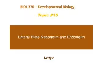
Chapter 15- Lateral mesoderm and endoderm
160 likes | 485 Views
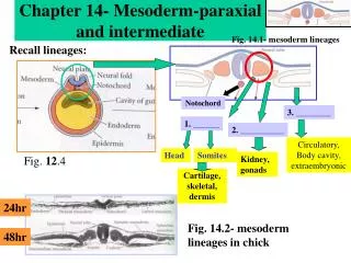
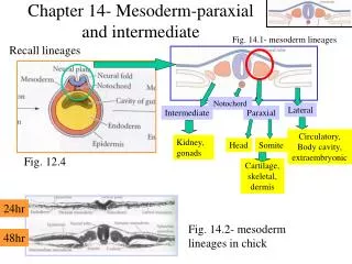
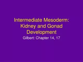
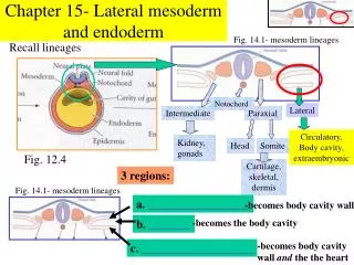
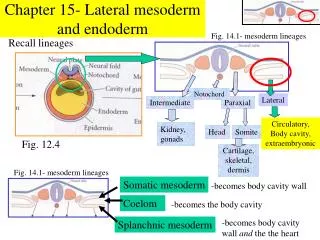
Fig. 14.1- mesoderm lineages. Recall lineages. Notochord. Lateral. Intermediate. Paraxial. Circulatory, Body cavity, extraembryonic. Kidney, gonads. Head. Somite. Fig. 12.4. Cartilage, skeletal, dermis. Chapter 15- Lateral mesoderm and endoderm. Fig. 14.1- mesoderm lineages.

Chapter 15- Lateral mesoderm and endoderm
E N D
Presentation Transcript
Fig. 14.1- mesoderm lineages Recall lineages Notochord Lateral Intermediate Paraxial Circulatory, Body cavity, extraembryonic Kidney, gonads Head Somite Fig. 12.4 Cartilage, skeletal, dermis Chapter 15- Lateral mesoderm and endoderm Fig. 14.1- mesoderm lineages Somatic mesoderm -becomes body cavity wall Coelom -becomes the body cavity -becomes body cavity wall and the the heart Splanchnic mesoderm
Lateral mesoderm How does the heart develop?? 25hr 26hr Fig. 15.3 Endocardium 1. Splanchnic mesoderm halves begin to merge 2. These cells differentiate into endocardium (heart lining and valve precursors and myocardium (heart muscles) 72hr 28hr 27hr Myocardium 3. Endocardium tubes fuse 4. Mycocardium fuses 5. Heart begins beating even while fusion is occurring
Blood vessel formation Lateral mesoderm 2 steps- vasculogenesis and angiogenesis Note: Blood vessels form independently of the heart, then link up Some background Info Constraints on blood vessel construction • Physiological- an organism must: • Obtain nourishment before the intestine develops • Use oxygen before there are lungs • Excrete wastes before there are kidneys Fig. 15.13- “extra” archs in mammal development • 2. Evolutionary- • Six pairs of aortic arches loop out- these enable primitive fish gills to oxygenate blood, but these serve no obvious purpose in mammals and birds. 3. Physical-Blood flows easier through large vessels, yet efficient diffusion requires small vessels and slow moving blood Solution- Large vessels branch into very small ones with overall more cumulative volume capacity
Blood vessel formation Lateral mesoderm 1. Vasculogenesis Fig. 15.14 Blood vessels and blood cells are intimately connected BMP Endothelial cells line blood vessels Angiogenic cell cluster (blood islands) Fig. 15.16 Primitive blood cells Mesenchyme Endothelial cells
Lateral mesoderm 1. Vasculogenesis Transcription factors in vasculogenesis 1. FGF2 is required for hemangioblast formation 2. VEGF is required for blood island and blood vessel formation VEGF is a target for tumor therapy “Tumors gotta eat” 3. Ang1 is required proper blood vessel formation (involved in communication between endothelial cell and smooth muscle)
Lateral mesoderm 2. Angiogenesis Definition- Remodeling and pruning of capillary beds, arteries and veins Note- Capillary networks of each organ arise within the organ itself, not from larger vessels! TGFb stabilizes capillary network VEGF plays key role PDGF recruits pericyte cells to ensure flexibility of capillaries
Lateral mesoderm 2. Angiogenesis Arteries vs. veins?? • Arteries have EphrinB2 in cell membranes • Veins have EphrinB2 receptor (called EphB4) in cell membranes Arterial (EphrinB2) Venous (EphB4) Fig. 15.17 Functions of the EphrinB2/EphB4 system • Ensure that arteries only link up with veins, not other arteries • Ensure capillary fusion only occurs with like cells (e.g. only arteries with arteries)
Lateral mesoderm 2. Angiogenesis Many organs make their own angiogenesis factors • Example- placenta Developing placenta secretes proliferin to promote angiogenesis, then later secretes proliferin-related protein to inhibit angiogenesis Angiogenesis plays key role in tumor development • A tumor must induce vascularization in order to enlarge • Hence, if use a drug that inhibits this angiogenesis, can possibly slow cure some cancers
Lateral mesoderm Development of Blood Cells Fig. 15.20 Stem cells – embryonic cells capable of producing many cell types, including other stem cells Largest population of stem cells is in the bone marrow “Committed” “Differentiating” “Differentiated” Stem Cell (CFU-M,L) B-cell lineage T-cell lineage
Lateral mesoderm Development of Blood Cells The stem cell (CFU-M,L) also gives rise to another cell lineage “Committed” “Differentiating” “Differentiated” Stem Cell (CFU-M,L) B-cell lineage Red blood cells Platelets Basophils T-cell lineage Myeloid precursor cell Eosinophils Neutrophils Fig. 15.21 Macrophage Note that this is the point of no return- cells are committed to a becoming only one cell type Paracrine factors that direct blood cell formation are termed “cytokines”
Lateral mesoderm Development of Blood Cells Blood development (hematopoiesis) occurs in two phases 1. Embryonic Angiogenic cell cluster (blood islands) • Occurs in blood islands in mesoderm near the yolk (recall fig. 15.16) • Supplies developing embryo with oxygen • BMP2 and 4 inhibit blood and blood vessel formation Fig. 15.16 • Transitory- disappear later in development Example- In mouse, stem cells originate in yolk sac, then later in AGM region 2. Definitive • Formed in nodes of mesoderm surrounding aorta (in a region called the aorta-gonad-mesonephros (AGM) region) • Lasts the lifetime of the individual Fig. 15.24
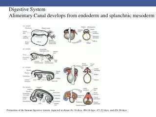
Endoderm Endoderm Recall Fig. 12.4 Embryonic endoderm gives rise two tubes Buds into Digestive tube (Esophagus,stomach,small intestine,colon) Liver, gallbladder, pancreas Primitive gut Endoderm Respiratory tube Lungs 1. Anterior endoderm – tissues are derived from pharyngeal arches Auditory cavities Tonsil walls Thymus (T-cell development) Parathyroid Pharyngeal arches Fig. 13.1 Lungs (sprout form base of forth arch)
2. Posterior endoderm Liver bud Stomach The hepatic diverticulum buds out form the foregut, then branches to form liver, pancreas and gall bladder Gall Bladder Pancreas (ventral) Pancreas (dorsal) The pancreas is actually formed by the fusion of two distinct buds (one ventral and one dorsal) Fig. 15.29
What directs formation of liver from the endoderm?? The notochord (and mesenchyme) produces factors that prevent liver induction The cardiac mesoderm secretes FGF that blocks the factors that inhibit liver induction Thus, FGF signals the proximal region of the endoderm to become liver Fig. 15.30
The respiratory tube • Lungs are one of the last organs to differentiate • Alveolar cells of the lung produce surfactant at 34 weeks gestation • Thus, a premature infant cannot breathe properly foregut Pharynx trachea Lung buds esophagus Fig. 15.31 Week 4 (humans)
Four problems of a land-dwelling egg Day 2 chick embryo Problem Solution 1. Desiccation Amnion secretes amnionic fluid into embryo 2. Gas Exchange Chorion exchanges gases 3. Nutrition Yolk duct supplies nutrients from blood vessels in yolk Day 9 chick embryo 4. Waste disposal- Allantois holds waste (vestigal in humans) Fig. 15.33