Download
1 / 57
570 likes | 599 Views
Explore the structures and functions of the male reproductive system, from external genitalia to internal organs, including testes, epididymis, vas deferens, and accessory glands. Learn about the anatomy of sperm and its pathway in the male system.

E N D
Anatomy of male and Female Reproductive Systems Dr. NabilKhouri
Objectives • Identify the structures related to the external genitalia • Identify the testis, its coverings, and tubules • Trace the entire course of the ductus deferens and identify its ampulla; • Identify the seminal vesicle and demonstrate the formation and course of the ejaculatory duct. • Identify the prostate gland and describe its subdivisions • Demonstrate the epididymis and its subdivisions. • Describe structure and function of the erectile bodies
Male Reproductive System What is the homologous structure of the scrotum in females?
External Genital Organs Penis Scrotum
Perineum in Lithotomy Position Urogenital trangle Anal trangle
Penis and the Superficial Perineal Pouch Glans penis Corpora spongiosa body Corpora cavernosa Crus of the penis roots Bullb of the penis
Penis The penis is the organ by which the sperm is introduced into the female. It contains spongy tissue that becomes turgid and erect when filled with blood.
Erectile Tissues • Corpus spongiosum– is the mass of spongy tissue whichsurrounds urethra and involves in erection by allowing rushing of blood into it • Corpus cavernosa– is one of a pair of songe-like regions of erectile tissue which contains most of the blood in the penis during penile erection
Coronal Section of the Penis Dorsal artery Superficial dorsal vein Dorsal nerve Deep dorsal vein Deep artery Superficial (Darto’s) fascia Corpora cavernosa (with tunica albuginea) Deep (Buck’s) fascia Corpora spongiosa (with tunica albuginea) Urethra
Sagittal View of the Penis Dorsal artery Superficial dorsal vein Deep dorsal vein Prepuce Dorsal nerve Corpora cavernosa (with tunica albuginea) Frenulum Corpora spongiosa (with tunica albuginea)
Urethra – a tube within the penis that conveys semen out of the body during ejaculation. Glans– the rounded, highly sensitive head of the penis. Prepuce – a fold of skin, covering the head of the penis.
Scrotum Median raphe A pouch of skin formed from the lower part of the abdominal wall. The scrotum keeps the testes at a temperature slightly cooler than body temperature.
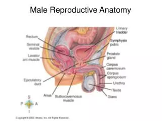
InternalGenital Organs Seminal Vesicles Prostate Gland Bulbourethral Glands Testis Epididymis Vas Deferens
The Scrotum and Testis Dartos muscle External spermatic fascia Cremaster muscle Internal spermatic fascia Vas deferens Epididymis Tunica vaginalis Testis
Testis The testes are the two-oval shaped male organs that produce sperm and hormone testosterone *. *Testosterone- the primary male sex hormone
Ductus deferens Head of epididymis Efferent ductules Septa Lobules Body of epididymis Tail of epididymis
Each testis is made of tightly coiled structures called seminiferous tubules. Among tubules are cells that produce testosterone.
Epididymis The epididymis is a tightly coiled tubes against the testicles. It acts as maturation and storage place for sperm. Adult human testicle with epididymis: A. Head of epididymis, B. Body of epididymis, C. Tail of epididymis, and D. Vas deferens
VasDeferens (Ductus Deferens) The vas deferens is a thin tube that starts from the epididymis to the urethra in the penis. They transport sperm from the epididymis in anticipation of ejaculation.
Accessory glands These glands produce nourishing fluids for the sperms that enter the urethra. Seminal Vesicles Prostate Gland Bulbourethral Glands
Seminal Vesicles produce a sticky yellowish fluid that contains fructose. Urinary bladder Ureter Vas deferens Seminal vesicle Ejaculatory duct Prostate The Seminal Vesicles are sac-like structures attached to the vas deferens at one side of the bladder.
Prostate Gland The Prostate Gland surrounds the ejaculatory ducts at the base of the urethra, just below the bladder. The Prostate Gland is responsible for making the production of semen, a liquid mixture of sperm cells, prostate fluid and seminal fluid.
Bulbourethral Glands(Cowper’s gland) The Bulbourethral Glands are two small glands located on the sides of the urethra just below the prostate gland. These glands produce a clear, slippery fluid that empties directly into the urethra.
Blood Supply of the Penis and Scrotum Common iliac artery Internal iliac artery External iliac artery Internal pudendal artery External pudendal artery Dorsal artery Deep artery Posterior scrotal artery Anterior scrotal artery
SPERM • Function: • To move and carry genetic information to the egg. • Structure: • Head: The large head region of the sperm that contains DNA. • Midpiece: The narrow middle part of the cell that contains mitochondria. • Tail: The wavelike motion of the flagellum propels the sperm forward.
REVIEW: The pathway of the sperm in the male reproductive system are the following: (arrange in chronological order)
Objectives • Describe the normal position and relationships of all organs of the reproductive tracts in both sexes and the role of each in reproductive processes. • Identify the vagina, and note the angle formed at its junction with the uterus • Identify the uterus and its subdivisions and demonstrate the continuity of its lumen with that of the uterine tubes and the vagina • Demonstrate the uterine tube and its subdivisions • Describe the broad ligament and differentiate its parts. • Identify the ovary and discuss the functional significance of its ligaments
Produce sex hormones Estrogen, Progesterone Produce egg (ova) Support & protect developing embryo Give birth to new baby Female Reproductive System
Vulva—which runs from the pubic area downward to the rectum. External genetalia • Labia majora or "greater lips" are the part around the vagina containing two glands (Bartholin’s glands) which helps lubrication during intercourse. • Labia minora or "lesser lips" are the thin hairless ridges at the entrance of the vagina, which joins behind and in front. In front they split to enclose the clitoris • The clitoris is a small pea-shaped structure. It plays an important part in sexual excitement in females.
The Female External Genitalia Mons pubis Clitoris Labia majora External urethral orifice Labia minora Vaginal orifice Hymen
Female External Genitalia and the Superficial Perineal Pouch Bulb of the Vestibule Greater vestibular gland (Bartholin’s gland) Perineal body Puborectalis Anus Pubococcygeus
The Female External Genitalia and the Superficial Perineal Pouch Clitoris Ischiocavernous muscle External urethral orifice Bulbospongiosus muscle Vaginal canal Superficial transverse perineal muscle
Female External Genitalia and the Deep Perineal Pouch Crus of the clitoris Clitoris Urethra Bulb of the vestibule Perineal membrane Vagina Greater vestibular gland Deep transverse perineal muscle
Sagital View of the Entire Female Reproductive System Fallopian tube Ovary Body of Uterus Cervix Vaginal canal Clitortis Labia minora Labia majora
A muscular, ridged sheath connecting the external genitals to the uterus. Functions as a two-way street, accepting the penis and sperm during intercourse Serving as the avenue of birth through which the new baby enters the world The Vagina Vesicouterine pouch Rectouterine pouch Pouch of Douglas Cul de sac Vagina Anterior and posterior fornices
The lower portion or neck of the uterus. The cervix is lined with mucus, known as cervical mucus Cervical mucus provides lubrication & sperm transport during sexual intercourse During ovulation secretion of cervical mucus increases in response to estrogen But when an egg is ready for fertilization, the mucus then becomes thin and slippery, offering a “friendly environment” to sperm The Cervix
The Cervix At the end of pregnancy The cervix acts as the passage through which the baby exits the uterus into the vagina. The cervical canal expands to roughly 50 times its normal width for the passage of the baby during birth
Pear-shaped muscular organ in the female reproductive tract. The fundus is the upper portion of the uterus where pregnancy occurs. The cervix is the lower portion of the uterus that connects with the vagina and serves as a sphincter to keep the uterus closed during pregnancy until it is time to deliver a baby. The uterus expands considerably during the reproductive process. The organ grows to from 10 to 20 times its normal size during pregnancy. Uterus
The main body consists of a firm outer coat of muscle (myometrium) and an inner lining of vascular, glandular material (endometrium). Uterus • The endometrium thickens during the menstrual cycle to allow implantation of a fertilized egg.
Uterus Fundus Endometrium Myometrium Body Perimetrium Isthmus Cervix
The endometrium is the innermost layer as a lining for the uterus During the menstrual cycle, the endometrium grows to a thick, blood vessel-rich, glandular tissue layer. This represents an optimal environment for the implantation of a blastocyst upon its arrival in the uterus. Endometrium
The endometrium is central, echogenic (detectable using ultrasound scanners), and has an average thickness of 6.7 mm. • During pregnancy, the blood vessels in the endometrium further increase in size and number, forming the placenta, • Placenta supplies oxygen and nutrition to the embryo & fetus. Endometrium
Also known as female gonads They produce eggs (also called ova) every female is born with a lifetime supply of eggs They also produce hormones: Estrogen & Progesterone Ovaries
Stretch from the uterus to the ovaries and measure about 8 to 13 cm in length. The ends of the fallopian tubes lying next to the ovaries feather into ends called fimbria Millions of tiny hair-like cilia line the fimbria and interior of the fallopian tubes. The cilia beat in waves hundreds of times a second catching the egg at ovulation and moving it through the tube to the uterine cavity. Fertilization typically occurs in the fallopian tube Fallopian tubes [uterine tubes]