Download
1 / 1
10 likes | 226 Views
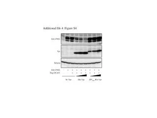
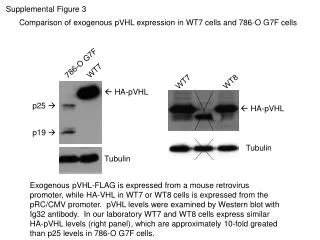
Supplemental Figure 3. Comparison of exogenous pVHL expression in WT7 cells and 786-O G7F cells. 786-O G7F. WT7. WT7. WT8. HA-pVHL. p25 . HA-pVHL. p19 . Tubulin. Tubulin.

E N D
Supplemental Figure 3 Comparison of exogenous pVHL expression in WT7 cells and 786-O G7F cells 786-O G7F WT7 WT7 WT8 HA-pVHL p25 HA-pVHL p19 Tubulin Tubulin Exogenous pVHL-FLAG is expressed from a mouse retrovirus promoter, while HA-VHL in WT7 or WT8 cells is expressed from the pRC/CMV promoter. pVHL levels were examined by Western blot with Ig32 antibody. In our laboratory WT7 and WT8 cells express similar HA-pVHL levels (right panel), which are approximately 10-fold greated than p25 levels in 786-O G7F cells.