Download
1 / 49
500 likes | 648 Views
Imaging Fascia. Renee Stenbjorn, MPA, LMT. My goal:. Utilize images to inform the student of the complex structural system of fascia. Your goal?. Historical Definition. Fascia: general description, a band of tissue.

E N D
Imaging Fascia Renee Stenbjorn, MPA, LMT
My goal: Utilize images to inform the student of the complex structural system of fascia. Your goal?
Historical Definition Fascia: general description, a band of tissue. General meaning: undifferentiated tissue surrounding a more specialized tissue. LANGEVIN, MD, H., HUIJING, PHD, P.. Communicating About Fascia: History, Pitfalls, and Recommendations.International Journal of Therapeutic Massage & Bodywork: Research, Education, & Practice, North America, 2, oct. 2009. Available at: <http://www.ijtmb.org/index.php/ijtmb/article/view/63>. Date accessed: 14 Jan. 2013.
Current understanding Forms of connective tissue: Loose & Dense Superficial & Deep Multiple- & Single-layered LANGEVIN, MD, H., HUIJING, PHD, P.. Communicating About Fascia: History, Pitfalls, and Recommendations.International Journal of Therapeutic Massage & Bodywork: Research, Education, & Practice, North America, 2, oct. 2009. Available at: <http://www.ijtmb.org/index.php/ijtmb/article/view/63>. Date accessed: 14 Jan. 2013.
Gross Anatomy Photography of anatomical structures: What is it? What do we learn?
"Old-School" Histology Gradations of pink, some purples, blues: Procedures: Create a specimen Mount on slides Put drops of solutions on slide for specific period of time. Rinse & mount. Put in a slide box.
"Old-School" Histology Put in a slide box.
What do we learn from histology? Highlights & differentiates: Cells, fibers & tissues Gives insight into cellular structures.
Histology of Connective Tissue types: Dense Irregular Abundant Collagen fibers, few cells. What type of tissues?
Histology of Connective Tissue types: Dense Regular Abundant Collagen fibers, arranged in same direction, f few cells. What type of tissues?
Histology of Connective Tissue types: Areolar Sparsely arranged fibers, not ordered in a predominant direction. Oftern refferd to as "Loose" but Dr. Langevin suggests this label is too mechanical in description
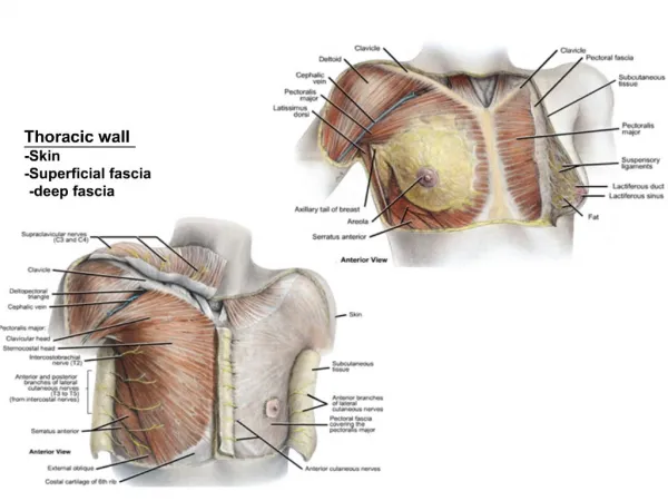
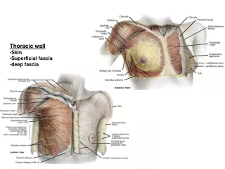
Specialized Fascial Structures: Superficial Fascia: aka: hypoderm, hypodermis, stratum subcutaneous, etc. Continuous with the dermis, directly underneath the skin
Specialized Fascial Structures: Deep Fascia
Specialized Fascial Structures: Muscular Fascial layers: Endo Peri Epi
Specialized Fascial Structures: Intermuscular Septa
Specialized Fascial Structures: Neurovascular Tract
Specialized Fascial Structures: Periosteum
How did histology help us differentiate: Regular vs. Irregular Fiber Arrangements Layers of fascia Structural differences
Imaging Cells & Fibers Immunohistochemistry: We can localize molecular structures rather than just cellular structures. Can measure DNA & RNA activity
What is different? Process of IHC Fluorescent labeling Co-localization of molecules
Dissecting a human brain... Dr. Liechnetz, MCV
Understanding Blots What do those dark spots mean?
Putting the theory to the test Can we find Collagen XVIII (18) in the brain? Using immunohistochemistry to locate structures.
Example: Cerebellum staining Collagen 18 found in Purkinje cells in the cerebellum!
Let's take a look: layers of fascia How images help us understand layers of tissue
Confocal imaging A method of converting IHC images into 3-D images that further our understanding of tissues.
3-D images Z Stack
The Neuromuscular Junction Understanding the relationship between nerve & muscle
The Extracellular Matrix What's in it? Why do we care? Key to hydration.
Sensory Mechanism Tensigrity architecture allows cells to sense mechanical changes. These changes result in control of cell function & tissue development Donald Ingber
Looking even deeper with Electron Microscopy Seeing the textures Understanding the structures
Imaging with Ultrasound From treatment to imaging: Principles in imaging with US. What can we see with US: what do these images tell us?
Video Imaging It's a living tissue.