Download

1 / 45
720 likes | 1.91k Views
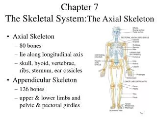
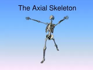
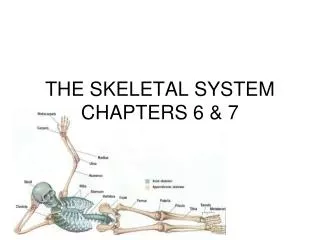
Chapter 7 The Skeletal System: The Axial Skeleton. Axial Skeleton 80 bones lie along longitudinal axis skull, hyoid, vertebrae, ribs, sternum, ear ossicles Appendicular Skeleton 126 bones upper & lower limbs and pelvic & pectoral girdles. 5 basic types of bones: long = compact

E N D
Chapter 7The Skeletal System:The Axial Skeleton • Axial Skeleton • 80 bones • lie along longitudinal axis • skull, hyoid, vertebrae, ribs, sternum, ear ossicles • Appendicular Skeleton • 126 bones • upper & lower limbs and pelvic & pectoral girdles
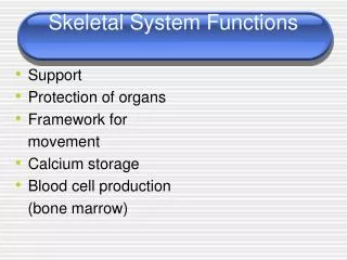
5 basic types of bones: long = compact short = spongy except surface flat = plates of compact enclosing spongy irregular = variable sesamoid = develop in tendons or ligaments (patella) Sutural bones = in joint between skull bones Types of Bones
Bone Surface Markings • Surface features-- rough area, groove, openings, process • Specific functions • passageway for blood vessels and nerves • joint formation • muscle attachment & contraction
Bone Surface MarkingsfromTable 7.2 • Foramen = opening • Fossa = shallow depression • Sulcus = groove • Meatus = tubelike passageway or canal • Condyle = large, round protuberance • Facet = smooth flat articular surface • Trochanter = very large projection • Tuberosity = large, rounded, roughened projection • Learning the terms found in this Table will simplify your study of the skeleton.
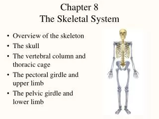
The Skull • 8 Cranial bones • protect brain & house ear ossicles • muscle attachment for jaw, neck & facial muscles • 14 Facial bones • protect delicate sense organs -- smell, taste, vision • support entrances to digestive and respiratory systems
The 8 Cranial Bones Frontal Parietal (2) Temporal (2) Occipital Sphenoid Ethmoid
Frontal Bone • Forehead, roof of orbits, & anterior cranial floor • Frontal suture gone by age 6 (metopic suture) • Supraorbital margin and frontal sinus
Parietal & Temporal Bones • Parietal • sides & roof of cranial cavity • Temporal • temporal squama • zygomatic processforms part of arch • external auditory meatus • mastoid process • styloid process • stylomastoid foramen(VII) • mandibular fossa (TMJ) • petrous portion (VIII)
Temporal and Occipital bones • Temporal • carotid foramen(carotid artery) • jugular foramen(jugular vein) • Occipital • foramen magnum • occipital condyles • external occipital protuberance attachment for ligamentum nuchae • superior & inferior nuchal lines
Sphenoid bone • Base of skull • Pterygoid processes are attachment sites for jawmuscles
Sphenoid in Anterior View • Body is a cubelike portion holding sphenoid sinuses • Greater and lesser wings • Pterygoid processes
Sphenoid from Superior View • Lesser wing & greater wing • Sella turcica holds pituitary gland • Optic foramen
Ethmoid Bone • Cranial floor, lateral nasal walls & nasal septum • Cribriform plate & olfactory foramina • Crista galli for attachment of membranes cover the brain
Ethmoid bone • Lateral masses contain ethmoid sinuses • Perpendicular plate is upper part of nasal septum • Superior & middle nasal concha or turbinates • filters & warms air
14 Facial Bones Nasal (2) Maxillae (2) Zygomatic (2) Mandible (1) Lacrimal (2) Palatine (2) Inferior nasal conchae (2) Vomer (1)
Maxillary bones • Floor of orbit, floor of nasal cavity or hard palate • Maxillary sinus • Alveolar processes hold upper teeth • Cleft palate is lack of union of maxillary bones
Zygomatic Bones • Cheekbones • Lateral wall of orbit along with sphenoid • Part of zygomatic arch along with part of temporal
Lacrimal and Inferior Nasal Conchae • Lacrimal bones • part of medial wall of orbit • lacrimal fossa houses lacrimal sac • Inferior nasal concha or turbinate (not part of ethmoid) Inferior Nasal Conchae
Palatine & Vomer • Palatine • L-shaped : one end is back part of hard palate, other end is part of orbit (see previous picture) • Vomer • posterior part of nasal septum
Mandible • Body, angle & rami • Condylar & coronoid processes • Alveolar processes for lower teeth • Mandibular & mental foramen
Sutures • Lambdoid suture unites parietal and occipital • Sagittal suture unites 2 parietal bones
Sutures • Coronal suture unites frontal and both parietal bones • Squamous suture unites parietal and temporal bones
Paranasal Sinuses • Paired cavities in ethmoid, sphenoid, frontal and maxillary • Lined with mucous membranes and open into nasal cavity • Resonating chambers for voice, lighten the skull • Sinusitis is inflammation of the membrane (allergy)
Fontanels of the Skull at Birth. • Dense connective tissue membrane-filled spaces(soft spots) • Unossified at birth but close early in a child's life. • Fetal skull passes through the birth canal. • Rapid growth of thebrain during infancy
Foramina of the Skull • Table 7.4 describes major openings of skull • In which bone would you find the following and what is their function? • foramen magnum • optic foramen • mandibular foramen • carotid canal • stylomastoid foramen
Bones of the Orbit • Roof is frontal and sphenoid • Lateral wall is zygomatic and sphenoid • Floor is maxilla, zygomatic and sphenoid • Medial wall is maxilla, lacrimal, ethmoid and sphenoid • Orbital fissures and optic foramen
Nasal Septum • Divides nasal cavity into left and right sides • Formed by vomer, perpendicular plate of ethmoid and septal cartilage • Deviated septum does not line in the midline • developmental abnormality or trauma
Hyoid Bone • U-shaped single bone • Articulates with no other bone of the body • Suspended by ligament and muscle from skull • Supports the tongue & provides attachment for tongue, neck and pharyngeal muscles
Vertebral Column • Backbone or spine built of 26 vertebrae • Five vertebral regions • cervical vertebrae(7) in the neck • thoracic vertebrae ( 12 ) in the thorax • lumbar vertebrae ( 5 ) in the low back region • sacrum (5, fused) • coccyx (4, fused)
Intervertebral Discs • Between adjacent vertebrae absorbs vertical shock • Permit various movements of the vertebral column • Fibrocartilagenous ring with a pulpy center
Normal Curves of the Vertebral Column • Primary curves • thoracic and sacral are formed during fetal development • Secondary curves • cervical if formed when infant raises head at 4 months • lumbar forms when infant sits up & begins to walk at 1 year
Typical Vertebrae • Body • weight bearing • Vertebral arch • pedicles • laminae • Vertebral foramen • Seven processes • 2 transverse • 1 spinous • 4 articular • Vertebral notches
Intervertebral Foramen & Spinal Canal • Spinal canal is all vertebral foramen together • Intervertebral foramen are 2 vertebral notches together
Typical Cervical Vertebrae (C3-C7) • Smaller bodies • Larger spinal canal • Transverse processes • shorter • transverse foramen for vertebral artery • Spinous processes of C2 to C6 often bifid • 1st and 2nd cervical vertebrae are unique • atlas & axis
Atlas & Axis (C1-C2) • Atlas -- ring of bone, superior facets for occipital condyles • nodding movement at atlanto-occipital joint signifies “yes” • Axis -- dens or odontoid process is body of atlas • pivotal movement at atlanto-axial joint signifies “no”
Thoracic Vertebrae(T1-T12) • Larger and stronger bodies • Longer transverse & spinous processes • Facets or demifacets on body for head of rib • Facets on transverse processes (T1-T10) for tubercle of rib
Lumbar Vertebrae • Strongest & largest • Short thick spinous & transverse processes • back musculature
Sacrum • Union of 5 vertebrae (S1 - S5) by age 30 • median sacral crest was spinous processes • sacral ala is fused transverse processes • Sacral canal ends at sacral hiatus • Auricular surface & sacral tuberosity of SI joint
Coccyx • Union of 4 vertebrae (Co1 - Co4) by age 30 • Caudal or epidural anesthesia during delivery • into sacral hiatus anesthetize sacral & coccygeal nerves • sacral and coccygeal cornu are important landmarks
Thorax • Bony cage flattened from front to back • Sternum (breastbone) • Ribs • 1-7 are true ribs (vertebrosternal) • 8-12 are false ribs (vertebrochondral) • 11-12 are floating • Costal cartilages • Bodies of the thoracic vertebrae.
Sternum • Manubrium • 1st & 2nd ribs • clavicular notch • Body • costal cartilages of 2-10 ribs • Xiphoid • ossifies by 40 • CPR position • abdominal mm. • Sternal puncture • biopsy
Ribs Fracture at site of greatest curvature. • Increase in length from ribs 1-7, thereafter decreasing • Head and tubercle articulate with facets • Body with costal groove containing nerve & blood vessels • Intercostal spaces contain intercostal muscles
Rib Articulation • Tubercle articulates with transverse process • Head articulates with vertebral bodies
Herniated (Slipped) Disc • Protrusion of the nucleus pulposus • Most commonly in lumbar region • Pressure on spinal nerves causes pain • Surgical removal of disc after laminectomy
Clinical Problems • Abnornal curves of the spine. • scoliosis (lateral bending of the column) • kyphosis (exaggerated thoracic curve) • lordosis (exaggerated lumbar curve) • Spina bifida is a congenital defect • failure of the vertebral laminae to unite • nervous tissue is unprotected • paralysis