Overview of Nervous System: Structure, Neurons, and Functions
Learn about the components of the nervous system, neuron structure, and functions of neurons. Explore the divisions of the nervous system and the role of neuroglia in supporting neurons.

Overview of Nervous System: Structure, Neurons, and Functions
E N D
Presentation Transcript
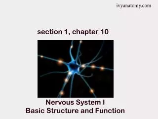
ivyanatomy.com section 1, chapter 10 Nervous System I Basic Structure and Function
The nervous system is divided into 2 subdivisions The Central Nervous System (CNS) consists of the brain and spinal cord. • The Peripheral Nervous System (PNS) • Consists of 12 pairs of cranial nerves and 31 pairs of spinal nerves • Nerves may be motor (efferent), sensory (afferent), or both (mixed)
Divisions of the PNS • The Somatic Nervous System is under voluntary control • Somatic motor controls skeletal muscles • Somatic sensory relays info regarding touch, pressure, and pain to the brain • The Autonomic Nervous System is under involuntary control • autonomic motor controls smooth muscles, cardiac muscles, • and glands • autonomic sensory relays visceral info regarding pH, blood • gasses, etc. to the brain
Figure 10.2. (a) overview of nervous system. CNS is grey, PNS is yellow. (b) CNS receives sensory input from PNS, and sends motor output to PNS. Somatic division of PNS is under voluntary control, while the autonomic division is under involuntary control.
The Autonomic Nervous System (ANS) is further divided into two branches. • The Sympathetic branch • prepares the body to respond to a stressful situation. • “Fight or Flight” Response • The Parasympathetic branch • Maintains normal body activities at rest • “Resting and Digesting”
Cells of the Nervous System • Neurons • Integrate, regulate, and coordinate body functions • Functions • Receive information - sensory • Conduct impulses - motor • Connect neurons - integrative • Neuroglia (glia = “glue”) • Neuroglia provide neurons with nutritional, structural, and functional support
Neurons Neurons vary in shape and size 3 Components of a neuron Dendrites receive impulse Call body (soma) Axon – transmits the impulse away from the cell body
Dendrites • Dendrites conduct information to the soma. • A cell may have a few dendrites, many dendrites, or no dendrites. • Dendritic Spines are additional contact points on some dendrites that increase the number of synapses possible by a neuron Cell Body – Soma Contains organelles such as the nucleus Mitochondria, Golgi Apparatus, etc. The rough ER is often called chromatophilic substance (Nissl Bodies)
Axon Axon Hillock is a specialized part of the soma that connects to the axon. The axon hillock is often called the Trigger Zone because action potentials begin here. Each neuron has only 1 axon, but it may divide into several branches, called collaterals The end of the axon is called the axon terminaland it enlarges into a synaptic knob (bouton)
Axon Microtubules called neurofibrils support long axons and aid in axonal transport (transport of biochemicals between the soma and the axon terminal)
Myelination of Axons The myelin sheath is a thick fatty coating of insulation surrounding the axon that greatly enhances the speed of impulses. Myelination of axons occurs differently in the PNS than in the CNS. Myelination of axons in the PNS. • Schwann Cells form the myelin sheath in the PNS. • They wrap around the axons in a jelly-roll fashion to form a thick layer of fatty insulation. • The cytoplasm and the nucleus are pushed to the outermost layer, forming the neurolemma • Schwann cells are separated by gaps, called Nodes of Ranvier.
Schwann cells still surround the axons of unmyelinated neurons, but they do not form the myelin sheath. Unmyelinated axons with a Schwann cell. A myelinated axon with a Schwann Cell.
Myelination of Axons in CNS Within the CNS the myelin sheath is formed by Oligodendrocytes. 1 Oligodendrocyte may form the myelin sheath of several axons. Gray and White Matter A mass of myelinated axons in the CNS forms the white matter. A mass of cell bodies (which are unmyelinated) along with unmyelinated axons form the gray matter. end of section 1, chapter 10
ivyanatomy.com Chapter 10, Section 2 Neurons and Neuroglia
Structural Classification of Neurons • A multipolar neuron contains many dendrites and 1 axon. • Includes most neurons in the brain and motor neurons • A bipolar neuron contains 1 dendrite and 1 axon • Includes some sensory neurons such as photoreceptors and olfactory neurons • A unipolar neuron contains a single process extending from the soma • Example includes the cells of the dorsal root ganglion • Peripheral Process – conducts information from PNS • Central Process – conducts information to CNS
Functional Classification of Neurons • An afferent or sensory neuron conducts information from the PNS to CNS • Dendrites may act as receptors (eyes, ears, touch) • Most afferent neurons are unipolar, and some are bipolar • An efferent or motor neuron conducts impulses from CNS to PNS • Voluntary Control – in somatic nervous system • Involuntary control – in autonomic nervous system An interneuron or association neuron is located completely within the CNS. Interneurons link neurons together in the CNS, and they also connect sensory neurons to motor neurons.
Functional Classification of Neurons Figure 10.7. Neurons classified by their functions. Sensory, Motor, and Interneurons.
Neuroglia in the CNS are different from those in the PNS Neuroglia in CNS • Astrocytes “star-shaped” attach blood vessels to neurons. • Astrocytes aid in metabolism, strengthen synapses, and participate in the blood-brain-barrier • Ependymal cells line the central canal of the spinal cord and the ventricles of the brain. • Ependyma regulate the composition of cerebral spinal fluid (CSF)
Neuroglia in CNS • Microgliaare normally small cells, but they enlarge into macrophages during an infection. • Phagocytize bacteria and cell debris • Oligodendrocytes form the myelin sheath in the CNS
Figure 10.8. Types of neuroglia in the CNS. Neuroglia compose half of the brain’s volume
Neuroglia of the PNS • Schwann Cells form the myelin sheath in the PNS. • Satellite Cells support clusters of cell bodies, called ganglia in PNS.
Disorders of Neuroglia Multiple Sclerosis (MS) The immune system attacks neurons in the CNS, destroying the myelin sheath of neurons. The damaged myelin sheath is replaced with Connective tissue, leaving behind scars (scleroses) Scars block the transmission of underlying neurons, so muscles no longer receive stimulation and begin to whither (atrophy). End Section 2, Chapter 10
ivyanatomy.com section 3, chapter 10 The Synapse And Membrane Potential
Synaptic Transmission Synaptic transmission is the mechanism that transmits a signal from the pre-synapticneuron to the post-synaptic neuron. An action potential causes the release of neurotransmitters from the presynaptic cell that diffuse across the synapse and bind to the postsynaptic cell.
Steps involved in Synaptic Transmission A nerve impulse (action potential) travels down the axon to the axon terminal. The action potential opens calcium channels causing calcium to diffuse into the synaptic knob. The calcium influx triggers the release of neurotransmitters from synaptic vesicles into the synapse. The neurotransmitters diffuse across the synapse and bind to receptors on the post-synaptic cell Some neurotransmitters are inhibitory whereas others are excitatory, so the post-synaptic cell may be stimulated or it may be inhibited depending on the neurotransmitter.
Cell Membrane Potential • The cell membrane is usually polarized (charged) • Inside the membrane is negatively charged relative to outside the membrane • Polarization is due to unequal distribution of ions across the membrane • Polarization is maintained by a series of ion pumps and channels
Factors that maintain the cell membrane potential 1. Sodium/Potassium (Na+/K+) pump • The sodium/potassium pump actively transports 3Na+ out of the cell, and 2K+ into the cell. • It creates a high extracellular [Na+] and a • high intracellular [K+] • requires ATP • The Na+/K+pump only contributes a small amount (-5mV) to the membrane potential.
Factors that maintain the cell membrane potential • Non-gated potassium channels “K+ leak channels” • The cell membrane has many K+ leak channels, but only a few Na+ leak channels • K+ continually leaks out of the cell, making the inside of the cell more negative.
Factors that maintain the cell membrane potential Figure A. The sodium-potassium pumps transports sodium out of the cell, while transporting potassium into the cell. Figure B. Leak channels allow some of the potassium to leak out of the cell, contributing to the positively charged extracellular fluid.
Factors that maintain the cell membrane potential The distribution of ions across the membrane creates a membrane potential (electrical gradient). For a neuron at rest the membrane potential is -70mV inside the cell. This is the Resting Membrane Potential RMP = -70mV inside the cell.
Factors that change the cell membrane potential Gated ion channels open and close in response to a stimulus.
Gated Ion Channels • 1. Mechanically-Gated Channels • Open or close in response to physical stress. • Touch, hearing, vibrations, ect. • 2. Ligand-Gated Ion Channels • Open or close in response to a ligand (neurotransmitter, hormone, or other molecule) • Includes ACh receptors on motor endplates • 3. Voltage-Gated Ion Channels • Open or close in response to small changes in the membrane potential (millivolts = mV) • Voltage-gated Na+ channels open when membrane potential reaches -55mV.
Gated Ion Channels Figure 10.15b. Ligand-gated Na+ channels (blue) open in response to neurotransmitters. Voltage-gated Na+ channels (pink) open in response to changes in membrane potential. End of section 3, chapter 10
Chapter 10, Section 4 Graded and Action Potentials
Changes in Membrane Potential • Resting Membrane Potential (RMP) for a neuron= -70mV • Membrane potential of a cell at rest • Environmental stimuli cause changes in membrane potential by opening gated ion channels • Ligand-gated ion channels • Voltage-gated ion channels • Other-gated ion channels • (respond to mechanical, temperature, or other stimulus) If membrane potential becomes more negative, it has hyperpolarized e.g. A membrane potential of -100mV is hyperpolarized If membrane potential becomes less negative, it has depolarized e.g. A membrane potential of -60mV is depolarized
Local Potential Changes • Graded Potentials • Local changes in membrane potential (usually occurs at dendrites) • Magnitude of response is proportional to stimulus • Graded potentials summate (add together) • Graded potentials generate action potentials If a graded potential reaches threshold stimulus (-55mV), it results in an action potential
Summation of Graded Potentials Summation of graded potentials my occur by: Spatial Summation – stimulating multiple dendrites Temporal Summation – Stimulating a dendrite at a high frequency Combined – stimulating multiple dendrites at a high frequency • Graded Potentials are summed together at the Axon Hillock “Trigger Zone” • If summation of graded potentials reaches threshold stimulus (-55mV), an action potential is initiated at the axon hillock.
Figure 10.15. (a) a subthreshold depolarization will not result in an action potential. (b) Summation of graded potentials may reach threshold stimulus, initiating an action potential at the trigger zone. The action potential begins when voltage-gated Na+ channels open at the trigger zone.
3 Phases of an Action Potential • Depolarization Phase • Voltage-gated Na+ channels open at -55mV (threshold stimulus) • Na+ diffuses into cell • Repolarization Phase • Voltage-gated K+ channels open at +30mV • K+ rushes out of the cell repolarizing the membrane • Na+ channels close • Hyperpolarization Phase • The slower voltage-gated K+ channels remain open briefly, resulting in a slight hyperpolarization (-90mV). Figure 10.17. An oscilloscope records and action potential 1 2 3
Action Potential Figure 10.16(a) At rest, the membrane is polarized (RMP = -70mV). Sodium is mostly outside the cell and potassium is within the cell. Figure 10.16(b) When a stimulus reaches threshold stimulus (-55mV), voltage-gated Na+ channels open. With Na+ channels open, sodium rapidly diffuses into the cell, depolarizing the membrane up to +30mV.
Action Potential Figure 10.16(c) When the membrane reaches +30mV, voltage-gated K+ channels open an quickly repolarize the membrane. Sodium channels also close at this point. Following an action potential, Na+/K+ pumps work to actively reestablish the Na+ and K+ concentration gradients.
Action Potential Propagation Once initiated an action potential is propagated along the entire axon at full strength. It does not weaken. Figure 10.18 An action potential in one region, depolarizes the adjacent region to threshold stimulus (-55mV). Once the adjacent region reaches threshold stimulus, it triggers another action potential. The second action potential causes depolarization in its adjacent region, triggering yet another action potential. This sequence continues all the way to the end of the axon at full strength.
All-Or-None Response • All-or-none response • Action potentials occur completely, or they do not occur at all. • An action potential occurs whenever a stimulus of threshold intensity • or above is applied to a neuron. • Greater stimulation does not produce a stronger impulse • (although a greater stimulation will produce more impulses per second)
Refractory Period Refractory Period: For a brief period following an action potential, a threshold stimulus will not trigger another action potential. • Absolute Refractory Period • no new action potentials can be produced • Occurs while the membrane is changing in sodium permeability • Between the depolarization and repolarization phases • Relative Refractory Period • Action potential can be generated with a high intensity stimulus • Occurs while membrane is reestablishing its resting membrane potential • Lasts from the hyperpolarization phase, until RMP is reestablished End of Chapter 10, Section 4
ivyanatomy.com section 5, chapter 10 Impulse Conduction & Neurotransmitters
Impulse Conduction Myelinated axons conduct impulses differently than unmyelinated axons. Unmeylinated Axon generates a series of action potentials along the entire axon The impulse is slow (travels at 1 mile/hour) Myelinated Axon Myelin is an electrical insulator and prevents action potentials along myelinated portions of the axon. Action potentials are generated only at the Nodes of Ranvier The impulse travels through the myelinated portions by electrical conduction. The impulse is fast (travels at 285 miles/hour!)