Download
1 / 32
400 likes | 633 Views
Explore various cardiovascular imaging modalities and stress testing techniques, including ECG, echocardiography, SPECT, CT angiography, MRI, and invasive procedures. Learn the general principles, indications, pros, and cons of each method.

E N D
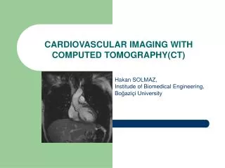
Cardiovascular imaging Cyril Štěchovský Dept. of Cardiology Motol University Hospital
Cardiovascular imaging • General principles of stress tests • Stress ECG • Transthoracic echocardiography (TTE) • Dobutamine stress ECHO • Transesophageal echocardiography (TOE) • Perfusion scintigraphy (SPECT) • CT angiography (CTA) / coronarography (CTC) • Cardicac MRI (CMRI) • Invasive coronary angiography (ICA) / peripheral angiography • Invasive hemodynamic measurement
General principles of stress tests • Ischemia testing • Maximal workload testing (VO2max) • Contractile reserve testing • Viability testing • Before the test: • Assess the pretest probability • Know sensitivity and specificity • Are there therapeutic consequences?
Ischemia testing SPECT, perfusion MRI, perfusion CT Dobutamine ECHO, stress MRI Anatomy – coronary stenosis: ICA CTC Stress ECG Chest pain
Ischemia testing • What type of test and how to induce ischemia? • Would stress ECG be diagnostic (LBBB, RBBB, WPW, stimuation, ST-segment changes: cardiomyopathy, LV hypertrophy, aortic stenosis, digoxin)? • Is exercise on ergometer or treadmill possible? • Shall I use vasodilating agent (dipyridamol, regadenoson, adenosine, nitroglycerine)? • Shall I use inotropic agent (dobutamine)?
Stress ECG • Pros: Cheap, available, fast, non-invasive • Cons: low sensitivity, many non-diagnostic findings, does not localize ischemia • Aim: adequate workload (W/kg, age dependent), at least submaximal heart rate 0.8x(220-age) • Ischemia = horizonatl or descending ST-depressions at least 1mm in two leads • Abnormal: hypertensive hyperreaction > 250/115 mmHg, drop in blood pressure, nsVT, new Afib, claudications, chest pain, dyspnea
Stress ECHO • Dobutamine up to 40ug/kg/min • Pros: higher sensitivity, assessment of contractile reserve • Cons: requires good imaging window and high expertise, side effects of dobutamine
Stress ECHO • Ischemia testing = new wall motion abnormality • Viability testing = contraction of previously akinetic segment, increase of LVEF • Low-flow low-gradient aortic stenosis = increase of LVEF and gradient on AV
SPECT • i.v. Radioactive agent (Tc-MIBI, thallium) that binds to membranes in cardiocytes • Detection of gamma radiation • Measurement of relative LV perfusion • Pros: Quantification and localization of ischemia, good specificity, viability??? • Cons: Intermediate sensitivity, high radiation dose
CT-coronarography • i.v. iodine contrast agent, beta-blocker to slow HR and nitroglycerine do dilatate coronary arteries, prospective ECG gating during end-diastole • Pros: superb sensitivity, fast, additional anatomic information (coronary anomalies, heart chambers, valves, aortic root, lungs, pulmonary artery), calcium score • Cons: lower specificity for ischemia, poor performance in Afib, requires very good CT and expert reader • Additional (experimental) modalities: perfusion CT, CT-FFR
Cardiac MRI • i.v. Gadolinium contrast agent, prospective imaging planes • Pros: best for myocardial diseases, LV and RV volumes, masses, function and viability testing and for congenital heart diseases • Cons: Expansive, time consuming, expet reader, perfusion and stress wall motion MRI are rarely available, does not show coronary arteries
Coronary angiography • Access site via radial, femoral or brachial artery with seldinger technique (needle, wire, sheath). • Selective engagement of left and right coronary ostia with performed shaped catheter (4-6F = 1,33-2mm) • In selected cases ventriculography and aortography with pigtail catheter • Pros: Golden standard for coronary stenosis assessment, additional intravascular imaging and preassure measurement (IVUS, OCT, NIRS, FFR), direct PCI • Cons: Expansive, bleeding and contrast nefropathy
Fractional flow reserve (FFR) • Introduction of FFR wire or catheter via guiding coronary catheter distal to the stenosis • Measurement of distal preassure (Pd) and aortic preassure (Pa) after administration of intracoronary adenosine • FFR = Pd/Pa • Golden standard of ischemia detection (FFR<0.80)
Hemodynamic measurement • Direct invasive measurement of pressures and saturation and assessment of cardiac output with thermodilution or O2 consuption (Fick) • CVP – RA – RV – PA- PCWP (right side) • Aorta – LV (left side) • CO/CI • Calculated: TPG, diastolic pulmonary gradient, PVR/PVRI, SVR/SVRI
Hemodynamic measurement Vilém Ganz 1919-2009, Czech cardiologist and the co-inventor of the Swan-Ganz catheter. Swan-Ganz catheter
Thank you for You attention! Without imaging a doctor is blind as a mole.