Download
1 / 25
250 likes | 413 Views
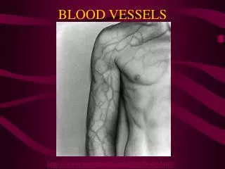
Biology 221 Anatomy & Physiology II. TOPIC 3 Circulatory System – Blood Vessels. Chapter 20 pp. 718-747. E. Lathrop-Davis / E. Gorski / S. Kabrhel. Blood Vessel Functions and Types. Function conduits for blood

E N D
Biology 221 Anatomy & Physiology II TOPIC 3Circulatory System – Blood Vessels Chapter 20 pp. 718-747 E. Lathrop-Davis / E. Gorski / S. Kabrhel
Blood Vessel Functions and Types • Function • conduits for blood • separate systemic and pulmonary systems provide more efficient delivery of oxygen and nutrients, removal of wastes • Major Types of Vessels • Arteries – carry blood away from the heart • Capillaries – sites of exchange of materials between blood and tissues • Veins – return blood to heart
Blood Vessel Histology Three layers of blood vessel wall: • Tunica interna (tunica intima) inner layer • endothelium (simple squamous epithelium) • subendothelial layer (areolar CT) • Tunica media – middle layer • varying amounts of dense connective tissue • smooth muscle • vasomotor tone • vasoconstriction • vasodilation http://www.usc.edu/hsc/dental/ghisto/cv/d_1.html Fig. 20.1, p. 719
Blood Vessel Histology • Tunica externa – outermost layer • connective tissue • nerve fibers, lymphatic vessels • vasa varsorum – blood vessel system in tunica externa of larger blood vessels (e.g., elastic arteries) http://www.usc.edu/hsc/dental/ghisto/cv/d_2.html http://www.usc.edu/hsc/dental/ghisto/cv/d_5.html
Elastic (Conducting) Arteries • Aorta and its major branches • Functions: • carry blood rapidly away from heart toward capillary beds • decrease blood pressure fluctuations during heart beat: • expand during ventricular systole, which decreases systolic pressure; and • recoil during ventricular diastole, which maintains pressure on blood
Elastic (Conducting) Arteries • Structure: • large diameter, large lumen • thick walls • lots of elastic fibers (elastin) • vaso vasorum (see previous slide) http://www.usc.edu/hsc/dental/ghisto/cv/d_2.html
Muscular (Distributing) Arteries • Account for most of the named arteries • Function: deliver blood to organs; control flow to organs • Structure: • internal diameter smaller than elastic arteries • thick tunica media with lots of smooth muscle http://www.usc.edu/hsc/dental/ghisto/cv/d_12.html
Arterioles • Function: distribute blood to tissues within organs major controller of blood flow into capillaries • Structure: • branch and become smaller • walls’ thickness decreases as they near capillaries • consists of endothelium and scattered smooth muscle cells near capillaries http://www.usc.edu/hsc/dental/ghisto/cv/d_12.html
Capillaries • Function: sites of exchange between blood and tissues • Structure: consist of tunica interna only • some with scattered pericytes (smooth muscle cells) • Three structural types: • continuous capillaries • fenestrated capillaries • sinusoids http://www.usc.edu/hsc/dental/ghisto/cv/d_25.html Fig. 20.3, p. 724
Continuous Capillaries • Endothelial cells continuous • cells may be held together by tight junctions • intercellular clefts are gaps in tight junctions • tight junctions are continuous in brain (blood-brain barrier) Fig. 20.3, p. 724 http://www.usc.edu/hsc/dental/ghisto/cv/d_25.html
Fenestrated Capillaries • Some endothelial cells have pores; most pores covered with membrane • Very permeable – allow even large substances to pass • Found in small intestine, some endocrine glands, kidney glomeruli Fig. 20.3, p. 724 http://www.usc.edu/hsc/dental/ghisto/cv/d_26.html
Sinusoids • Large irregular lumens slows blood flow • Walls fenestrated or incompletely lined with endothelial cells • in liver, endothelium is discontinuous where macrophages (Kupffer cells) form part of vessel wall • in spleen, phagocytes on outside of endothelial lining extend processes into lumen • Few tight junctions allow large molecules (e.g., proteins) to pass through • Located in liver, spleen, bone marrow, lymphoid tissue, some endocrine glands Fig. 20.3, p. 724 http://www.usc.edu/hsc/dental/ghisto/cv/d_34.html
Capillary Beds • Many capillary branches from arteriole • Microcirculation = flow of blood from arteriole to venule through capillary bed • Metarteriole - thoroughfare channel • fast, direct connection between arteriole and venule • terminal arteriole metarteriole thoroughfare channel venule • by-passes capillary bed when tissue is inactive Fig. 20.4, p. 725
Capillary Beds: True Capillaries • sites of exchange between blood and tissues • branches of metarteriole • rejoin to thoroughfare channel • precapillary sphincter controls blood movement into capillary bed • amount of blood entering depends on gross needs of body (vasomotor nervous control) and local needs of tissue (local chemical cues) Fig. 20.4, p. 725
Post-capillary Venules • Function: collect blood from capillary beds • Structure: • formed by union of capillaries • leaky endothelium with few pericytes • White blood cells (WBCs) abundant http://www.usc.edu/hsc/dental/ghisto/cv/d_36.html
Veins • Functions: • carry blood under low pressure back toward heart • act as blood reservoirs = capacitance vessels; • ~ 65% of body’s blood is in veins http://www.usc.edu/hsc/dental/ghisto/cv/d_39.html
Veins • Structure: • gradually increase in size and thickness • all 3 tunics present, but thinner than in arteries of corresponding size (external diameter) • little smooth muscle or elastin • relatively thicker tunica externa • valves prevent backflow • varicose veins - blood pools because valves fail causing venous walls to expand • Phlebitis – inflammation of vein http://www.usc.edu/hsc/dental/ghisto/cv/d_39.html
Venous Sinuses • Function: collect blood under low pressure • Structure: • flattened veins with walls of endothelium only • supported by surrounding tissues • Examples: coronary sinus; dural sinuses
Vascular Anastomoses • Arterial anastomoses form collateral channels to maintain flow in case of blockage • Examples: • brain - circle of Willis • around joints • abdominal organs • heart • Arteriovenous anastomoses • metarteriole thoroughfare channel • Venous anastomoses • vein to vein • more common
Circulatory Patterns General pattern: Ventricles of heart elastic arteries muscular arteries arterioles capillaries venules veins atria of heart See Table 20.1, p. 721
Circulatory Patterns Two main systems: Pulmonary circulation: • right ventricle pulmonary trunk R/L pulmonary arteries alveolar capillaries of the lungs R/L pulmonary veins left atrium • takes deoxygenated blood to lungs for exchange of gases • Systemic circulation: • left ventricle aorta arteries capillaries of body tissues superior and inferior venae cavae right atrium • takes oxygenated blood to tissues, removes wastes See Fig. 20.2, p. 720
Special Circulatory Patterns: • Hepatic Portal System - carries venous blood from intestines, pancreas, stomach, spleen to liver; Fig. 20.27, p. 771 • Hypophyseal Portal System - carries blood with regulatory hormones from hypothalamus to anterior pituitary; Fig. 17.5, p. 617
Special Circulatory Patterns: • Coronary Circulation (covered with heart) - carries oxygen-rich blood to heart and removes oxygen-poor blood; Fig. 19.7, p. 690 • Cerebral Circulation - carotid and vertebral arteries carry oxygen-rich blood to brain and jugular veins drain oxygen-poor blood from brain (details in lab); Fig. 20.20. p. 755 Fig. 20.25, p. 767
Special Circulatory Patterns: • Fetal Circulation - Fig. 29.13, p. 1136 • by-pass developing lungs • ductus arteriosus – carries blood from pulmonary trunk to aorta • foramen ovale – allows blood to flow from right to left atrium • Gas & material exchange at placenta • umbilical arteries carry deoxygenated blood high in wastes to placenta • umbilical vein brings oxygenated blood high in nutrients from placenta • What do these fetal structures become? • What happens if they fail to close?
Vessel Disorders • Atherosclerosis:blood vessel walls are abnormally thick and narrowed and less compliant • Occlusive coronary atherosclerosis: narrowing of coronary arteries due to accumulation of cholesterol; often lead to ischemia and infarct • Arteriosclerosis:late stage of atherosclerosis • Aneurysm: localized dilation or out-pouching of a blood vessel or a cardiac chamber; rupture often leads to severe bleeding