Physiological Psyc
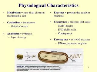
Physiological Psyc. Ch.2. Cells of the Nervous System. The nervous system is composed of two types of cells, neurons and glia . Neurons. Cells which receive and transmit information to other cells. The human brain contains approximately 100 billion neurons.


Physiological Psyc
E N D
Presentation Transcript
Physiological Psyc Ch.2
Cells of the Nervous System The nervous system is composed of two types of cells, neurons and glia
Neurons • Cells which receive and transmit information to other cells. • The human brain contains approximately 100 billion neurons. • The finding that the brain like the rest of the body is composed of individual cells was demonstrated by Santiago Ramón y Cajal in the late 1800s.
Neurons • Membrane (plasma membrane): Composed of two layers of fat molecules; this membrane allows some small uncharged chemicals to flow both into and out of the cell. Protein channels allow a few charged ions to cross the membrane, however most chemicals are unable to cross.
Neurons • Nucleus: The structure that contains the chromosomes. • Mitochondrion: The structure that provides cell with energy. Requires fuel and oxygen to function.
Neurons • Ribosomes: Site of protein synthesis in the cell. • Endoplasmic reticulum: A network of thin tubes that transports newly synthesized proteins to other locations. Ribosomes may be attached.
Structure of a Neuron • Most neurons contain four major components: dendrites, cell body, axon, and presynaptic terminal. Small neurons may lack axons and well-defined dendrites.
A motor neuron: Conduct impulses to muscles and glands from the spinal cord. • A sensory neuron (receptor neurons): Sensitive to certain kinds of stimulation (e.g., light, touch, etc.).
Dendrites: Branching fibers which extend from the cell body and get narrower at their end. The dendrite’s surface is lined with specialized synaptic receptors, at which the dendrite receives information from other neurons. • Dendritic spines: Short outgrowths found on some dendritic branches.
Cell body (soma): Contains the nucleus, ribosomes, mitochondria, and other structures found in most cells. • Axon: A long, thin fiber (usually longer than dendrites) which is the information-sending part of the neuron, sending an electrical impulse toward other neurons, glands, or muscles.
Myelin sheath: Insulating covering found on some vertebrate axons. • Presynaptic terminal (end bulb or bouton): Swelling at the tip of the axon. Part of the neuron which releases chemicals that cross the junction between one neuron and the next.
Neurons may have any number of dendrites, but are limited to no more than one axon (which may have branches).
Afferent axons: Brings information into a structure. • Efferent axons: Sends information away from a structure. • Interneurons (intrinsic neuron): Entirely located within a single structure of the nervous system.
Neurons vary enormously in size, shape, and function. • A neurons function is closely related to its shape. • A neuron’s shape is plastic (changeable) as new experiences can modify the shape of a neuron.
Glia • Glia are the other major component of the nervous system. Glia have many different functions but they do not transmit information like neurons. • A 10:1 ratio of glia to neurons exists in the brain.
Astrocytes: A type of glia that absorbs chemicals released by axons and later returns those chemicals back to the axon to help synchronize the activity of neurons. Also, astrocytes remove waste products, particularly those created after neurons die.
Microglia: Very small cells that remove waste material as well as viruses, fungi and other microorganisms. • Oligodendrocytes: A type of glia that builds the myelin sheaths around certain neurons in the brain and spinal cord.
Schwann cells: A type of glia that builds the myelin sheaths around certain neurons in the periphery of the body. • Radial glia: Type of astrocyte. Guides the migration of neurons and the growth of axons and dendrites during embryonic development.
The Blood-Brain Barrier • The mechanism that keeps most chemicals out of the vertebrate brain. • The blood-brain barrier is needed because the brain lacks the type of immune system present in the rest of the body. The area postrema is not protected by the blood brain barrier and monitors blood chemicals that could not enter other parts of the brain. This area triggers nausea and vomiting.
The blood-brain barrier works because endothelial cells forming the walls of the capillaries in the brain are tightly joined blocking most molecules from passing. In the rest of the body the endothelial cells are separated by large gaps.
Small uncharged molecules (e.g., oxygen and carbon dioxide) and molecules that can dissolve in the fats of the capillary wall can cross passively (without using energy) through the blood-brain barrier.
An active transport system (a protein-mediated process that uses energy) exists to pump necessary chemicals, such as glucose, through the blood-brain barrier.
Nourishment of Vertebrate Neurons • Almost all neurons depend on glucose (a simple sugar) for their nutrition. • Neurons rely on glucose so heavily because glucose is practically the only nutrient that crosses the blood-brain barrier in adults. Ketones can also cross but are in short supply.
A thiamine (vitamin B1) deficiency leads to an inability to use glucose, which could lead to neuron death and a condition called Korsakoff's syndrome (a disorder marked by severe memory impairment).
The Resting Potential • The membrane of a neuron maintains an electrical gradient (a difference in electrical charge between the inside and outside of the cell). • In the absence of any outside disturbance (i.e., at rest), the membrane maintains an electrical polarization (i.e., a difference in electrical charge between two locations) that is slightly more negative on the inside relative to the outside. This difference in electrical potential or voltage is known as the resting potential.
The resting potential is measured by very thin microelectrodes. A typical resting membrane potential is –70 millivolts (mV).
The neuron membrane has selective permeability which allows some molecules to pass freely (e.g., water, carbon dioxide, oxygen, etc.) while restricting others. Most large molecules and ions cannot cross the membrane. A few important ions cross through protein channels.
During the resting potential, potassium and chloride channels (or gates) remain open along the membrane which allows both ions to pass through; sodium gates remain closed restricting the passage of sodium ions.
Sodium-potassium pump • a protein complex found along the neuron membrane which transports three sodium ions outside of the cell while also drawing two potassium ions into the cell; this is an active transport mechanism (requires energy to function). The sodium-potassium pump causes sodium ions to be more than ten times more concentrated outside than inside.
When the membrane is at rest, two forces work on sodium ions: • The electrical gradient • Concentration gradient
The electrical gradient: opposite electrical charges attract, thus sodium (which is positively charged) is attracted to the negative charge inside the cell.
Concentration gradient :(difference in distribution of ions between the inside and the outside of the membrane): Sodium is more concentrated outside the membrane than inside and is thus more likely to enter the cell than to leave it. • Given that both the electrical and concentration gradients tend to move sodium into the cell, sodium would be expected to quickly enter the cell. However, when the membrane is at rest sodium channels are closed.
Potassium ions are subject to the same two forces, however, the forces are in opposition to each other. Potassium ions are positively charged so the electrical gradient tends to move potassium in, but since potassium is concentrated on the inside of the cell the concentration gradient causes potassium to flow out of the cell.
The Action Potential • Hyperpolarization (increased polarization): Occurs when the negative charge inside the axon increases (e.g., -70mV becomes -80mV). • Depolarization (reduced polarization towards zero): Occurs when the negative charge inside the axon decreases (e.g., -70mV becomes -55mV).
Threshold of excitation (threshold): The level that a depolarization must reach for an action potential to occur. • Action potential: A rapid depolarization and slight reversal of the usual membrane polarization. Occurs when depolarization meets or goes beyond the threshold of excitation.
When the potential across an axon membrane reaches threshold, voltage-activated (membrane channels whose permeability depends on the voltage difference across the membrane) sodium gates open and allow these ions to enter) this causes the membrane potential to depolarize past zero to a reversed polarity (e.g., -70mV becomes +50mV at highest amplitude of the action potential).
When the action potential reaches its peak, voltage-activated sodium gates close, and potassium ions flow outside of the membrane due to their high concentration inside the neuron as opposed to outside. Also, the electrical gradient is now pushing potassium flow outward.
A temporary hyperpolarization (membrane potential below the resting potential) occurs before the membrane returns to its normal resting potential (this is due to potassium gates opening wider than usual, allowing potassium to continue to exit past the resting potential).
After the action potential, the neuron has more sodium and fewer potassium ions for a short period (this is soon adjusted by the sodium-potassium pumps to the neuron's original concentration gradient).
Local anesthetic drugs (e.g., Novocain, Xylocaine, etc.) block the occurrence of action potentials by blocking voltage-activated sodium gates (preventing sodium from entering a membrane). • General anesthetics (e.g., ether and chloroform) cause potassium gates to open wider, allowing potassium to flow outside of a neuron very quickly.
All-or-none law: The size, amplitude, and velocity of an action potential is independent of the intensity of the stimulus that initiated it. If threshold is met or exceeded an action potential of a specific magnitude will occur, if threshold is not met, an action potential will not occur.
Refractory period • Absolute refractory period: Sodium gates are incapable of opening; hence, an action potential cannot occur regardless of the amount of stimulation. • Relative refractory period: Sodium gates are capable of opening, but potassium channels remain open; a stronger than normal stimulus (i.e., exceeding threshold) will initiate an action potential.
Propagationof the Action Potential • The action potential begins at the axon hillock (a swelling located where the axon exits the cell body). • The action potential is regenerated due to sodium ions moving down the axon, depolarizing adjacent areas of the membrane.
The action potential moves down the axon by regenerating itself at successive points on the axon. • The refractory periods prevent the action potentials from moving in the opposite direction (i.e., toward the axon hillock).
Myelin Sheath & Saltatory Conduction • Myelin: an insulating material composed of fats and proteins found on some vertebrate axons. Myelin greatly increases the speed of propagation • Myelinated axons: axons covered with a myelin sheath.
Nodes of Ranvier: Short unmyelinated sections on a myelinated axon. • Saltatory conduction: The "jumping" of the action potential from node to node. • Some diseases including multiple sclerosis destroy the myelin along axons; loss of the myelin sheath slows down or prevents the propagation of action potentials.
Local Neurons • neuron with short dendrites • a short (if any) axon
Local neurons do not produce action potential but communicate with their closest neighbors using graded potentials (membrane potentials that vary in magnitude and do not follow the all-or-none law). Graded potentials get smaller as they travel.