Download
1 / 40
400 likes | 588 Views
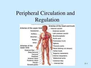
19. The Cardiovascular System: Blood Vessels: Part A. Blood Vessels. Delivery system of dynamic structures that begins and ends at the heart Arteries: carry blood away from the heart; oxygenated except for pulmonary circulation and umbilical vessels of a fetus

E N D
19 The Cardiovascular System: Blood Vessels: Part A
Blood Vessels • Delivery system of dynamic structures that begins and ends at the heart • Arteries: carry blood away from the heart; oxygenated except for pulmonary circulation and umbilical vessels of a fetus • Capillaries: contact tissue cells and directly serve cellular needs • Veins: carry blood toward the heart
Arterial system Venous system Large veins (capacitance vessels) Heart Elastic arteries (conducting vessels) Large lymphatic vessels Lymph node Muscular arteries (distributing vessels) Lymphatic system Small veins (capacitance vessels) Arteriovenous anastomosis Lymphatic capillary Sinusoid Arterioles (resistance vessels) Postcapillary venule Terminal arteriole Metarteriole Thoroughfare channel Precapillary sphincter Capillaries (exchange vessels) Figure 19.2
Structure of Blood Vessel Walls • Arteries and veins • Tunica intima, tunica media, and tunica externa • Lumen • Central blood-containing space • Capillaries • Endothelium with sparse basal lamina
Tunica intima Valve • Endothelium • Subendothelial layer Internal elastic lamina Tunica media (smooth muscle and elastic fibers) External elastic lamina Tunica externa (collagen fibers) Lumen Vein Lumen Artery Capillary network Basement membrane Endothelial cells Capillary (b) Figure 19.1b
Tunics • Tunica intima • Endothelium lines the lumen of all vessels • In vessels larger than 1 mm, a subendothelial connective tissue basement membrane is present
Tunics • Tunica media • Smooth muscle and sheets of elastin • Sympathetic vasomotor nerve fibers control vasoconstriction and vasodilation of vessels
Tunics • Tunica externa (tunica adventitia) • Collagen fibers protect and reinforce • Larger vessels contain vasa vasorum to nourish the external layer
Elastic (Conducting) Arteries • Large thick-walled arteries with elastin in all three tunics • Aorta and its major branches • Large lumen offers low-resistance • Act as pressure reservoirs—expand and recoil as blood is ejected from the heart
Muscular (Distributing) Arteries and Arterioles • Distal to elastic arteries; deliver blood to body organs • Have thick tunica media with more smooth muscle • Active in vasoconstriction
Arterioles • Smallest arteries • Lead to capillary beds • Control flow into capillary beds via vasodilation and vasoconstriction
Capillaries • Microscopic blood vessels • Walls of thin tunica intima, one cell thick • Pericytes help stabilize their walls and control permeability • Size allows only a single RBC to pass at a time
Capillaries • In all tissues except for cartilage, epithelia, cornea and lens of eye • Functions: exchange of gases, nutrients, wastes, hormones, etc.
Arterial system Venous system Large veins (capacitance vessels) Heart Elastic arteries (conducting vessels) Large lymphatic vessels Lymph node Muscular arteries (distributing vessels) Lymphatic system Small veins (capacitance vessels) Arteriovenous anastomosis Lymphatic capillary Sinusoid Arterioles (resistance vessels) Postcapillary venule Terminal arteriole Metarteriole Thoroughfare channel Precapillary sphincter Capillaries (exchange vessels) Figure 19.2
Capillaries • Three structural types • Continuous capillaries • Fenestrated capillaries • Sinusoidal capillaries (sinusoids)
Continuous Capillaries • Abundant in the skin and muscles • Tight junctions connect endothelial cells • Intercellular clefts allow the passage of fluids and small solutes • Continuous capillaries of the brain • Tight junctions are complete, forming the blood-brain barrier
Pericyte Red blood cell in lumen Intercellular cleft Endothelial cell Basement membrane Tight junction Pinocytotic vesicles Endothelial nucleus (a) Continuous capillary. Least permeable, and most common (e.g., skin, muscle). Figure 19.3a
Fenestrated Capillaries • Some endothelial cells contain pores (fenestrations) • More permeable than continuous capillaries • Function in absorption or filtrate formation (small intestines, endocrine glands, and kidneys)
Pinocytotic vesicles Red blood cell in lumen Fenestrations (pores) Endothelial nucleus Intercellular cleft Basement membrane Endothelial cell Tight junction (b) Fenestrated capillary. Large fenestrations (pores) increase permeability. Occurs in special locations (e.g., kidney, small intestine). Figure 19.3b
Sinusoidal Capillaries • Fewer tight junctions, larger intercellular clefts, large lumens • Usually fenestrated • Allow large molecules and blood cells to pass between the blood and surrounding tissues • Found in the liver, bone marrow, spleen
Endothelial cell Red blood cell in lumen Large intercellular cleft Tight junction Nucleus of endothelial cell Incomplete basement membrane (c) Sinusoidal capillary. Most permeable. Occurs in special locations (e.g., liver, bone marrow, spleen). Figure 19.3c
Capillary Beds • Interwoven networks of capillaries form the microcirculation between arterioles and venules • Consist of two types of vessels • Vascular shunt (metarteriole—thoroughfare channel): • Directly connects the terminal arteriole and a postcapillary venule
Capillary Beds • True capillaries • 10 to 100 exchange vessels per capillary bed • Branch off the metarteriole or terminal arteriole
Blood Flow Through Capillary Beds • Precapillary sphincters regulate blood flow into true capillaries • Regulated by local chemical conditions and vasomotor nerves
Vascular shunt Precapillary sphincters Thoroughfare channel Metarteriole True capillaries Terminal arteriole Postcapillary venule (a) Sphincters open—blood flows through true capillaries. Terminal arteriole Postcapillary venule (b) Sphincters closed—blood flows through metarteriole thoroughfare channel and bypasses true capillaries. Figure 19.4
Venules • Formed when capillary beds unite • Very porous; allow fluids and WBCs into tissues • Postcapillary venules consist of endothelium and a few pericytes • Larger venules have one or two layers of smooth muscle cells
Veins • Formed when venules converge • Have thinner walls, larger lumens compared with corresponding arteries • Blood pressure is lower than in arteries • Thin tunica media and a thick tunica externa consisting of collagen fibers and elastic networks • Called capacitance vessels (blood reservoirs); contain up to 65% of the blood supply
Vein Artery (a) Figure 19.1a
Pulmonary blood vessels 12% Systemic arteries and arterioles 15% Heart 8% Capillaries 5% Systemic veins and venules 60% Figure 19.5
Veins • Adaptations that ensure return of blood to the heart • Large-diameter lumens offer little resistance • Valves prevent backflow of blood • Most abundant in veins of the limbs • Venous sinuses: flattened veins with extremely thin walls (e.g., coronary sinus of the heart and dural sinuses of the brain)
Vascular Anastomoses • Interconnections of blood vessels • Arterial anastomoses provide alternate pathways (collateral channels) to a given body region • Common at joints, in abdominal organs, brain, and heart • Vascular shunts of capillaries are examples of arteriovenous anastomoses • Venous anastomoses are common
Physiology of Circulation: Definition of Terms • Blood flow • Volume of blood flowing through a vessel, an organ, or the entire circulation in a given period • Measured as ml/min • Equivalent to cardiac output (CO) for entire vascular system • Relatively constant when at rest • Varies widely through individual organs, based on needs
Physiology of Circulation: Definition of Terms • Blood pressure (BP) • Force per unit area exerted on the wall of a blood vessel by the blood • Expressed in mm Hg • Measured as systemic arterial BP in large arteries near the heart • The pressure gradient provides the driving force that keeps blood moving from higher to lower pressure areas
Physiology of Circulation: Definition of Terms • Resistance (peripheral resistance) • Opposition to flow • Measure of the amount of friction blood encounters • Generally encountered in the peripheral systemic circulation • Three important sources of resistance • Blood viscosity • Total blood vessel length • Blood vessel diameter
Resistance • Factors that remain relatively constant: • Blood viscosity • The “stickiness” of the blood due to formed elements and plasma proteins • Blood vessel length • The longer the vessel, the greater the resistance encountered
Resistance • Frequent changes alter peripheral resistance • Varies inversely with the fourth power of vessel radius • E.g., if the radius is doubled, the resistance is 1/16 as much
Resistance • Small-diameter arterioles are the major determinants of peripheral resistance • Abrupt changes in diameter or fatty plaques from atherosclerosis dramatically increase resistance • Disrupt laminar flow and cause turbulence
Relationship Between Blood Flow, Blood Pressure, and Resistance • Blood flow (F) is directly proportional to the blood (hydrostatic) pressure gradient (P) • If P increases, blood flow speeds up • Blood flow is inversely proportional to peripheral resistance (R) • If R increases, blood flow decreases: F = P/R • R is more important in influencing local blood flow because it is easily changed by altering blood vessel diameter