Download
1 / 1
10 likes | 118 Views
Study investigates the effects of Androstenediol on TLR3 signaling in murine macrophages. Results show AED does not enhance NF-κB pathway in TNF-α gene expression. Supported by NIH/NIDCR T32.DE014320-04.

E N D
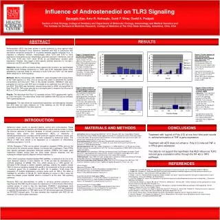
Influence of Androstenediol on TLR3 Signaling Byungdo Han, Amy R. Hufnagle, Scott F. Wray, David A. Padgett Section of Oral Biology, College of Dentistry and Department of Molecular Virology, Immunology and Medical Genetics and The Institute for Behavioral Medicine Research, College of Medicine at The Ohio State University, Columbus, Ohio, USA ABSTRACT RESULTS Androstenediol (AED) has been shown to confer protection to mice against lethal infections with influenza A virus. Moreover, treatment with AED in conjunction with vaccination enhanced production of circulating antibody against influenza virus. In response to influenza infection, macrophages utilize TLR3 and TLR7 to drive expression of the genes for pro-inflammatory cytokines and type I interferons. Like all TLRs, both TLR3 and TLR7 utilize NF-κB for pro-inflammatory cytokine gene expression. However, they trigger a more specific antiviral response (i.e, the type I interferons) via IRF3 and IRF7 respectively. Objectives: Due to AED's protective effects against viral infections, we hypothesized that AED would increase transcription of TLR3- and TLR7- dependent genes. After establishing a general model for activation of both TLR3 and TLR7, we first tested AED's influence on TLR3 signaling. Methods: Murine macrophage cells, RAW264.7, were stimulated with varying doses of the TLR3 agonist (i.e. Poly (I:C)) at various time points to determine the optimal concentration and time point. Then, in the optimal condition, RAW264.7 cells were pretreated for one hour with AED and co-treated for an additional hour with Poly (I:C) and AED. Total RNA was harvested, reverse-transcribed to cDNA, and quantified by Real-Time PCR. TNF-α was selected as a surrogate gene to measure the influence of AED on TLR-3 induced NF-κB activity. Results: The data show that Poly (I:C) optimally induces TLR3 signaling with 1μg/mL at 1 hour time point. The data further reveal that treatment with AED did not enhance TNF-α gene expression. Conclusion: The data refute the experimental hypothesis and alternatively indicates that AED does not exert influence on TLR3 signaling via the NF-κB pathway. Supported by NIH/NIDCR T32 DE014320-04 Figure 1: Temporal Kinetics for Poly (I:C) stimulation. Previous studies indicated that 1µg/mL of poly (I:C) was optimal for stimulation of TLR-3 driven TNF-α gene expression in RAW264.7 macrophages. This figure illustrates the influence of 1µg/mL of poly (I:C) at multiple timepoints after stimulation. The data indicate that one-hour time point is sufficient for stimulation. Figure 2: Further analysis of dose and time on the activation of TLR-3 induced gene expression. This experiment focused on the concentrations of Poly(I:C) between 0.5 and 1.5µg/mL and on the time points between 0.5 and 1.5 hours. Data confirm that 1µg/mL of poly (I:C) at one-hour provides optimal condition of TLR-3 driven TNF-α gene expression in order to investigate the effects of AED. Figure 3: Effect of AED on TLR3 signaling as measured by TNF-α mRNA expression. This experiment evaluated the effect of AED on the TLR3 signaling pathway involving NF-κB. RAW264.7 cells were pre-treated with AED (1.0x10⁻⁷M) for one hour and subsequently co-treated with AED (1.0x10⁻⁷M) and Poly(I:C) (1.0µg/mL) for one hour. The data demonstrate that AED does not enhance poly (I:C) induced TNF-α. gene expression with is dependent upon NF-κB. Figure 4: Effect of AED on TLR3 signaling measured via IFN-β mRNA expression. This experiment evaluated the effect of AED on the TLR3 signaling pathway involving IRF3. RAW264.7 cells were pre-treated with AED (1.0x10⁻⁷M) for one hour and subsequently co-treated with AED (1.0x10⁻⁷M) and Poly(I:C) (1.0µg/mL) for one hour. The data demonstrate that AED does not enhance poly (I:C) induced IFN-β gene expression with is dependent upon IRF3. INTRODUCTION MATERIALS AND METHODS CONCLUSIONS Behavioral stress results in elevated plasma cortisol and corticosterone. These glucocorticoids possess powerfully anti-inflammatory effects that are known to impair the immune response to infectious diseases. In contrast, previous studies from this laboratory have shown that androstenediol (AED), a metabolite of dehydroepiandrosterone, provided antiviral protection against influenza, herpes, and coxsackie viruses in animals that had been subjected to behavioral stress. The effects of AED were shown to be dependent on a restoration of inflammatory cytokines expression (1,2,3,4). Toll-like Receptors (TLRs) act as pattern recognition receptors (PRRs) and are the tool by which the immune system detects it is infected with a pathogen. These PRRs bind specific pathogen associated molecular patterns (PAMPs) and trigger gene expression thereby driving both the initial innate immune response and the subsequent antigen-specific adaptive immune response (5). TLR3, which recognizes double-stranded RNA (dsRNA), is believed to be one of the most important sensors of viral infection (6). TLR3 can also be stimulated by a synthetic analogue of dsRNA, Poly (I:C), which serves as a TLR3 agonist (6). Activated TLR3 induces a distinct signaling pathway, utilizing the adaptor protein TRIF to induce a signaling cascade that results in the expression of the anti-viral type I interferon genes; this is meditated via IRF3 (8). In addition, TLR3 drives the expression of multiple inflammatory cytokine genes such as TNF-α; this effect is mediated by activation of NF-κB (9). TLR3 signaling is unique in that it does not utilize the adaptor protein MyD88, which is involved in all the other TLR signaling pathways. Because AED has demonstrated an ability to enhance antiviral protection, we hypothesized that AED would increase the expression of TLR3-induced inflammatory cytokine genes. In order to test this hypothesis, RAW264.7 macrophages were stimulated with 1µg/mL of poly (I:C) for one hour, and Real-Time PCR was employed to quantify the expression of TNF-alpha as an NF-κB-induced gene and IFN-beta as an IRF3/IRF7-induced gene. The data revealed that treatment with AED did not enhance TLR-3 driven gene expression. Cell Culture: Murine macrophage RAW264.7 (ATCC; Rockville, MD) were suspended in Roswell Park Memorial Institute (RPMI) media supplemented with 10% fetal bovine serum (FBS) and seeded in the 6-well plates 18 hoursbefore experimentation. A hemocytometer was used to count cells, and cultures were seeded with 106 cells in 1.0 mL. Immunostimulants: RAW264.7 cells were stimulated with varying concentrations of polyinosinic:polycytidylic acid (Poly I:C) (Sigma, St. Louis, MO) at various time points for the activation of TLR3 signaling pathway. Steroid preparation: AED was initially solubilized in 1:1 DMSO:EtOH as 1x10⁻²M and was serially diluted to achieve the concentration of 5x10⁻⁵M. The final concentration of DMSO:EtOH in the culture media was 2µg/mL. RNA Isolation: After stimulation, RNA was harvested by using Trizol Reagent (Invitrogen; Grand Island, NY). Spectrophotometric measurements at 260nm and 280nm wavelengths were used to determine the total concentrationand the purity of the isolated RNA samples. cDNA Synthesis: 1µg of RNA from each sample was to synthesize cDNA using utilizing a Promega kit with AMVReverse Transcriptase (Promega; Madison, WI). Real Time Polymerase Chain Reaction (PCR): Real Time PCR employed the ABI Prism 7700 Sequence DetectionSystem and the TaqMan kit (Applied Biosystems; Foster City, CA). Primers/Probes: Ribosomal RNA gene: 18S 18S Forward Primer: 5’-CTGTCTACTGAACTTCGGGGTGAT-3’ 18S Reverse Primer: 5’-GCTGGAATTACCGCGGCT-3’ Probe: 5’-TGCTGGCACCAGACTTGCCCTC-3’ Tumor Necrosis Factor Alpha gene: TNF-α TNF-α Forward Primer: 5’- CTGTCTACTGAACTTCGGGGTGAT –3’ TNF-α Reverse Primer: 5’-GGTCTGGGCCATAGAACTGATG-3’ Probe: 5’-ATGAGAAGTTCCCAAATGGCCTCCCTC-3’ Interferon-beta gene: IFN-beta: IFN-β IFN-β Forward Primer: 5’-TGAATGGAAAGATCAACCTCACCTA-3’ IFN-β Reverse Primer: 5’-CTCTTCTGCATCTTCTCCGTCA-3’ Probe: 5’-AGGGCGGACTTCAAGATCCCTATGGA-3’ Treatment with 1µg/mL of Poly (I:C) at one-hour time point results in optimal stimulation of TNF-α gene expression. Treatment with AED does not enhance Poly (I:C)-induced TNF-α or IFN-β gene expression. The data do not support the hypothesis that AED influences TLR3 induced gene expression either through the NF-κB or IRF3 pathways. REFERENCES 1. Padgett DA, Loria RM, Sheridan JF. Steroid hormone regulation of antiviral immunity. Ann N Y Acad Sci 2000;917:935-43. 2. Padgett DA, Sheridan JF. Androstenediol (AED) prevents neuroendocrine-mediated suppression of the immune response to an influenza viral infection. J Neuroimmunol 1999 Aug 3;98(2):121-9. 3. Daigle J, Carr DJ. Androstenediol antagonizes herpes simplex virus type 1-induced encephalitis through the augmentation of type I IFN production. J Immunol 1998 Mar 15;160(6):3060-6. 4. Loria RM, Padgett DA, Huynh PN. Regulation of the immune response by dehydroepiandrosterone and its metabolites. J Endocrinol 1996 Sep;150:S209-20. 5. Arancibia SA, Beltrán CJ, Aguirre IM, Silva P, Peralta AL, Malinarich F, Hermoso MA. Toll-like receptors are key participants in innate immune responses. Biol Res 2007;40(2):97-112. 6. Alexopoulou L, Holt AC, Medzhitov R, Flavell RA. Recognition of double-stranded RNA and activation of NF-kappaB by Toll-like receptor 3. Nature 2001 Oct 18;413(6857):732-8. 7. Marshall-Clarke S, Downes JE, Haga IR, Bowie AG, Borrow P, Pennock JL, Grencis RK, Rothwell P. Polyinosinic acid is a ligand for toll-like receptor 3. J Biol Chem 2007 Aug 24;282(34):24759-66. Epub 2007 Jun 14. 8. Taro Kawai, Shizuo Akira. Toll-like Receptor and RIG-I-like Receptor Signaling. Ann N Y Acad Sci 2008;1143:1-20. 9. Hiscott J, Nguyen T-LA, Arguello M, Nakhaei P, Paz S. Manipulation of the nuclear factor-kB pathway and the innate immune response by viruses. Oncogene 2006;25:6844-6867.