
Electron Microscopy
E N D
Presentation Transcript
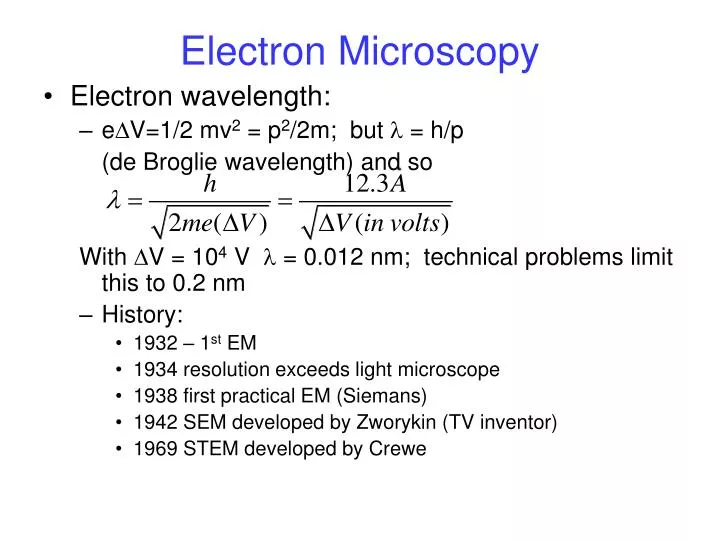
Electron Microscopy • Electron wavelength: • eDV=1/2 mv2 = p2/2m; but l = h/p (de Broglie wavelength) and so With DV = 104 V l = 0.012 nm; technical problems limit this to 0.2 nm • History: • 1932 – 1st EM • 1934 resolution exceeds light microscope • 1938 first practical EM (Siemans) • 1942 SEM developed by Zworykin (TV inventor) • 1969 STEM developed by Crewe
TEM Instrument Few um spot on sample Critical for aberrations Thermionic emission – 2400 K Accelerate through 40 – 100 kV Up to 1 MV in HV-EM Overall mag = 1000 – 300,000x
Sample Preparation • Grid with C film by vacuum deposition, or plastic film • Thin sections or liquid sample • Need to improve poor intrinsic contrast • 4 basic methods: • Replica – deposit thin layer of metal (Pt) and float off and look at replica • Freeze-etching – rapidly freeze and sublime water (to minimize damage) • Shadow – evaporate heavy metals (Au) • Negative stain with heavy metal salt (U)
Preparation of grids for EM work - Carbon-platinum replica: Coat with Pt, then C, then float film onto water and pick up on grid
Shadowing – vacuum deposition Kleinschmidt technique for DNA preparation for EM
Freeze-fracture + rotary shadowing human amnion cell
Details on SEM • Electron beam comes in off-axis at ~ 10 nm spot • Scanned across surface in raster pattern • Detectors sense a number of signals – backscattered e-, x-rays, fluorescence, also transmission • Signals are displayed as 2-D image on CRT • Samples must be coated and resolution is limited to ~ 10 nm – but depth of focus is extremely high
STEM • Record both elastic and inelastic scattering – ratio is characteristic of each element and so can do elemental analysis • Uses tightly focused beam (0.5 nm) and scans as in SEM, but at each point measures ratio of Ielastic/Iinelastic – very high resolution and elemental analysis