Download
1 / 79
820 likes | 1.19k Views
Week 9 c Chapter 39 Designing for Radiation Protection and Chapter 40 Radiation Protection. Many radiation protection devices and accessories are associated with a modern radiology service. Two of these common to all x-ray machines are: Protective Tube Housing The Control Panel

E N D
Week 9 c Chapter 39 Designing for Radiation Protection and Chapter 40 Radiation Protection • Many radiation protection devices and accessories are associated with a modern radiology service. Two of these common to all x-ray machines are: • Protective Tube Housing • The Control Panel • Many safety features are designed into the modern equipment.
Protective Tube Housing • Every x-ray tube must be contained within a protective housing to reduce leakage during operation. • The limit or leakage is less than 100 mR/hour.
Control Panel • The control panel must indicate the conditions of the exposure and positively indicate when the tube is energized. • Indications include: • mA, time or mAs • kVp • Focal Spot • A visible and audible signal that the tube is energized.
Source to Image Receptor Distance • A source to image receptor (SID) indicator must be provided. • It may be as simple as a ruler attached to the collimator. • It may be a ruler attached to the rails for horizontal and vertical tube movement. • It may be lights attached to the receptor or locks that prohibit exposure unless the SID is correct. • The SID indicator must be accurate to 2% of the SID.
Collimation • The unit should be equipped with a light localized, variable-aperture rectangular collimator. • Cone or diaphragms may be used in specialized machines. • Collimation must be accurate to 2% of the SID. • Some machines will have automatic collimation called PBL Positive Beam Limitation. It was required from 1974 to 1994 and still seen on many machines.
Beam Alignment • Each x-ray tube should have a mechanism to ensure that the tube is properly aligned to the image receptor. • It would do no good to align the light and beam if the film is not aligned.
Filtration • All general purpose x-ray units operated at 70 kVp or above must have at least 2.5 mm of Al filtration built into the unit. • Units operated between 50 and 70 kVp must have 1.5mm of Al filtration. • Below 50kVp 1.0 mm Al is required • Mammography tube have 30 µm of Mo or 60 µm of Rh.
Reproducibility • The out put radiation intensity should be constant from one exposure to another using the same factors. • Checked my making multiple exposures and observing the variation of intensity. • The variation can not exceed 5%.
Linearity • When adjacent mA stations are used such as 100 mA or 200 mA and the time is adjusted the intensity should remain the same. • When the time is changed, the change should be proportional. • The linearity of exposure should be within 5%.
Operator Shield • It should not be possible to make an exposure with the operator outside the operator shield or control booth. • The exposure button should be attached to the control panel and not on a long cord. • We use Dead man type switches that require two fingers to make the exposure.
Fluoroscopic Equipment • Since fluoroscopic equipment is beyond the chiropractic standards in California we will not cover all the protective devices in fluoroscopic machines. They include • Lead shields • Collimators • Exposure Controls • Bucky slot cover • Cumulative Timer
Design of Protective Barriers • When designing a radiographic room a number of factors must be addressed besides the architectural features. • Machine location • Range of movement of tube and tube direction • Use of adjoining rooms • Which floor you are on.
Design of Protective Barriers • Great attention must be given to the location of the equipment. • Often necessary to add a protective barrier lead sheets or other construction materials placed in the walls of the examination room. • They may even be required in the floor and rarely in the ceiling. • A medical physicist must be consulted to determine shielding requirement for the room.
Types of Radiation • The protective barriers must account for tube leakage, the primary beam and scatter radiation. • Primary radiation is most intense so it requires the greatest shielding.
Primary and Secondary Barriers • For erect radiography, the receptor is mounted to the wall. This wall and any wall that the primary beam can be directed are considered primary protective barriers. • The other walls are considered as secondary protective barriers. Tube leakage and the patient are the primary source of exposure to these walls. • The patient is the single most important source of secondary radiation. • The intensity of the scatter is about 0.1% of the primary beam at the patient.
Shielding materials • Usually lead sheets bonded to sheetrock or wood paneling are placed in the walls for primary protective barriers. • Such materials are available in various thickness as is specified to the architect and contractor in pounds per square foot. • Construction materials such as concrete block or brick can be used instead of lead.
Secondary Protective Barriers • The walls that are away from the tube direction are secondary barriers. The exposure comes from tube leakage and the patient. • Barriers designed to shield secondary radiation are called secondary protective barriers. They are always less thick than primary barriers. • Often lead is not required depending upon the distance from the tube. Steel, glass gypsum or wood may be adequate.
Control Booth • Ideally the control booth is located so the primary beam can not be directed toward the control booth. In this case, it is a secondary protective barrier. Four thicknesses of gypsum and ½” thick glass may be adequate. • If the tube can be directed toward the control booth as at the college, the wall facing the tube is a primary barrier.
Factors Affecting Barrier Thickness • Distance The distance from the source of radiation greatly impacts the thickness of the barrier. • Wall and floor mounted tube stands require more shielding on the attaching wall due to protection from tube leakage. • Ceiling suspended systems often have the table in the center of the room so no single wall is subjected to especially intense radiation.
Factors Affecting Barrier Thickness • Occupancy The use of the area protected is of principle importance. A closet or storeroom would need less shielding than an office or laboratory occupied 40 hours per week. • This reflects the time of occupancy factor that was established by the NCRP.
Factors Affecting Barrier Thickness • Controlled area: Design limits for controlled access areas are based upon the annual recommended dose limits of 5000 mrem/year. The weekly limit for exposure to personnel in the space is less than 100 mrem per week. • Controlled area are occupied by monitored radiology personnel and patients only. • Uncontrolled area: An uncontrolled area can be occupied by anyone. Exposure limits are based upon the public DL of 2 mrem/wk or 2.5 mrem in any hour.
Factors Affecting Barrier Thickness • Workload: The volume of radiographic examinations must be factored. Busy rooms require more shielding that infrequently used rooms. • This characteristic is called Workload (W) and units of measure of milliampere-minutes per week (mAmin/wk).
Factors Affecting Barrier Thickness • Use Factor: The percentage of time the tube is on and directed toward a particular wall is called the use factor. • For conventional x-raythe floor is rated as 1 and each wall as ¼. • Rooms with a wall Bucky has the wall behind the Bucky as 1. All others would be considered as zero for primary protection so they are secondary barriers. • Ceiling are almost always considered as secondary protective barriers.
Factors Affecting Barrier Thickness • kVp: The final consideration in the design of an x-ray protective barrier is the penetrability of the x-ray beam. kVp is the factor for penetration. • 100 kVp is used for general radiology rooms and 30 kVp for mammography.
The final result • Measurements taken outside the room always results in radiation levels far below the calculated exposure. The use of 100 kVp is high since most exposure are in the 75 kVp range. • The calculation are intended to result in exposure limits of 100 mrem/wk in the room and 2 mrem/wk outside the room. Rarely will the exposure exceed 1/10 of the DL.
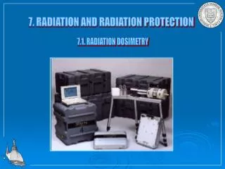
Radiation Detection and Measurement • There are instruments designed to detect radiation or to measure radiation or both. • Those designed to detect usually operated in a pulse or rate mode and are used to detect the presence of radiation. • They will chirp or tick when radiation is detected. • The response measurement is in mR/hr or R/hr.
Dosimeters • Units that measure the intensity of radiation are called dosimeters. They operate in the integrate mode where they accumulate the exposure and respond to the total exposure. • The response is in mR or R. • The earliest dosimeter was the film badge. It is still popular today though there are some newer technologies that have some better characteristics.
Dosimeters • Types of Dosimeters • Film Badge (photographic emulsion) • Gas filled detectors • Ion Chambers • Geiger-Muller Counter • Proportional Counters • Thermoluminescence dosimeter (TLD) • Optically stimulated dosimeter (OSL) • Scintillation detector
Film Badges • Limited range < 10 mR not measured. • Energy Dependent • Must be changed monthly • Popular for personnel monitoring • Must be worn with proper side to exposure. • Sensitive to heat never leave in a car. • Sensitive to humidity and water.
Gas-Filled Detectors • Can measure a wide range of radiation intensities from 1 mR/hr to several thousand r/hr. • Used to assay radionuclides in nuclear medicine.
Gas-Filled Detectors • The ionization of gas is the basis for gas filled detectors. • They are used as laboratory instruments and meters to detect very low radiation intensities.
Thermoluminescence Dosimeter (TLD) • Some materials glow when heated. This is referred to as thermoluminesecence. • Some materials will glow brightly after exposure to ionizing radiation and subsequently heated. • This is the principle of operation of the TLD. Discovered in the 1960 at the University of Wisconsin.
TLD • After irradiation, the TLD phosphor is placed on a special dish or plancet for analysis in the analyzer. • The analyzer is light tight and temperature controlled. • A PM tube is used to read the exposure.
TLD • Lithium fluoride is most commonly used TLD material. • It is relatively sensitive and can measure doses as low as 5 mrad with modest accuracy and at exposures greater then 10 rad is accurate to better than 5%. • Calcium Fluoride activated with manganese is more sensitive and can measure less than 1 mrad. Used for environmental monitoring.
TLD • TLD’s have the several benefits as personnel monitors compared to film badges. • They are reusable • More accurate • Not sensitive to heat or humidity • Can be changed less frequently i.e. quarterly
Optically Stimulated Luminescence (OSL) • Developed in the late 1990’s by Landauer, Optically Stimulated Luminescence uses aluminum oxide at the radiation detector. • The irradiation of Al2O3 stimulates some electrons to an excited state.
Optically Stimulated Luminescence (OSL) • During processing, laser light stimulates these electrons to an excited state and returning them to ground state with the production of light. • Works similar to TLD but far more accurate.
OSL • OSL is superior to TLD and Film Badges because: • Accurate to 1 mrad within +/-1 mrad. • Reanalysis is possible to confirm report. • Gain qualitative information about exposure conditions. • Wide range • Long Term stability
Scintillation Detectors • Scintillation detectors are the basis of nuclear medicine gamma cameras used for bone scans. • Scintillation detectors are used in computed tomography scanners. • Scintillation detectors are more sensitive than Geiger-Muller dosimeters.
Scintillation Detectors • The crystal emittes light proportional to the energy of the absorbed radiation. • The 50 keV exposure is totally absorbed and produces 50 units of light. • The second exposure produced 30 keV of Compton scatter and 20 units of light • A PM tube captures the light.
Occupational Radiation Exposure • Radiation dose is measured in units of rads (GyT). • Radiation exposure is measured in Roentgens (GyA). • When the radiation exposure is to a chiropractor, radiologic technologist or radiologist, the proper unit is rem (Sv).
REM (Sv) • The rem is the unit of effective dose and is used for radiation protection purposes. • The terms exposure, dose and effective dose have precise and different meanings but they are often used interchangeably in radiology because they have approximately the same numeric value. • The rem (Sv) identifies the biologic effectiveness of the radiation energy absorbed.
Recommended Dose • The recommended dose for radiology radiation workers is 50 mSv/year (5000 mrem/year). • Studies have shown that 88% receive less than 1 mSv/year (100 mrem/year). • 53% receive less than the detectible dose. • 0.05% receive more than 50 mSv/year.
Recommended Dose • Medical radiologist typically receive more radiation exposure than technologists due to fluoroscopy and being closer to the source of radiation.
Fluoroscopy • Fluoroscopy is the primary source of the highest occupational exposure. • Personnel in the room during the examination must wear protective aprons.
Fluoroscopy • The lead apron and how the tube is placed will have a great impact on exposure during fluoroscopy. • Radiologist and personnel doing interventional examinations should also wear extremity monitoring.
Computed Tomography • The operators area in computed tomography is safe. There is exposure in the scanner room so aprons should be worn when in the room during scanning.