Download
1 / 52
540 likes | 900 Views
Chapter 40 Radiation Protection Procedures. ALARA and Occupational Exposure. ALARA. ALARA stands for As Low As Reasonably Achievable. It is the basic principle of radiation protection procedures. There is much that we can do to keep exposure to the patient and the operator as low as possible.

E N D
Chapter 40 Radiation Protection Procedures ALARA and Occupational Exposure
ALARA • ALARA stands for As Low As Reasonably Achievable. It is the basic principle of radiation protection procedures. • There is much that we can do to keep exposure to the patient and the operator as low as possible. • The chiropractor is unique in the fact that you can perform radiography and refer your patients for other types of examinations.
Occupational Exposures • In radiologic technology, 95% of the occupational exposure comes from fluoroscopy and mobile radiography. • Neither would be used in your office so the worst case scenario is that you would receive 5% of the exposure that a technologist would receive.
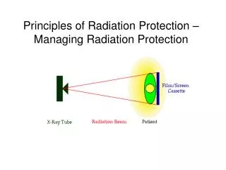
Occupational Exposures • During radiography, the operator should be behind a protective barrier. • These barriers are usually considered as secondary barriers so protection would be from tube leakage and scatter from the patient. The tube should never be pointed toward this type barrier. • If the barrier can have the tube angled toward the barrier. It must be a primary barrier. • Staying behind the barrier effectively eliminates the source of occupational exposure if the shielding is adequate.
Occupational Exposures • Medical Imaging Exposures • Fluoroscopy: All personnel will wear protective apron. If extremities get into the beam lead gloves can be worn. • The radiologist will usually be close to the machine during fluoroscopy so their exposure will be higher than that the technologist. Aprons between the Image intensifier and Bucky Slot covers reduce radiologist exposure. • The technologist should stand as far away from the table as possible during the exam and move closed only when necessary. • The radiologist will use short burst of exposure and keep the exposure time as short as possible. The 5 minute clock timer will alarm when 5 minutes of fluoroscopy has been used.
Occupational Exposures • Medical Imaging Exposures • Mobile radiography: • The technologist must wear a lead apron during mobile plain film or fluoroscopy examinations. • An apron must be assigned to each portable machine. • The exposure cord for portable radiographic machines must be 2 meters long to maximize distance from the tube during exposures.
Occupational Exposures • Radiology Ancillary Staff • Assuming the rooms are adequately shielded, the receptionist, file room and darkroom staff should not receive any occupational exposure. • Radiology ancillary staff should not be used to hold patients during radiography.
Occupational Radiation Monitoring • Occupational Radiation Monitoring is required if there is any likelihood that an individual will receive more than 1/10 of the recommended dose. • With just plain film radiography, monitoring may not be required as long as the operator stays in the control booth during all exposures. • There are some exams such as stress views of the ankle where the operator would be in the room with the patient. If this is done, monitoring would be necessary. • If the operator ever holds a patient monitoring would be necessary.
Occupational Radiation Monitoring • Occupational radiation monitoring offers no protection against exposure. It merely records the exposure received. • If needed, find a certified laboratory to process the dosimeters. • Types of monitors • Film badges • TLD • OSL
Film Badges • Film badges have been used since the 1940’s and are still used today. • Exposures below 10 mR are not measured on the film. • Filters along with the window in the badge allow estimation of the energy of the exposure. • The must be worn with the proper side to the front. • They are typically worn on the collar so they would remain outside the lead apron.
Film Badges • Advantages • Inexpensive • Easy to handle and process • Reasonably accurate • Disadvantages • Can not be reused • Sensitive to heat and humidity • Must be changes monthly
TLD • TLD has several advantages over film badges. • Not sensitive to heat or humidity • Measure exposures to 5mR More sensitive and accurate. • Can be changed quarterly instead of monthly • Disadvantages • Cost but changing badges less frequently than monthly eliminates cost problem.
Optically Stimulated Luminescence • All of the advantages of the TLD over film badges plus: • Can be re-read to confirm exposure • More accurate than TLD
Where to wear the monitor • The whole body badge is typically worn at collar level so it can be outside the lead apron. • Fetal monitoring badges used during pregnancy are worn at waist level under the apron. • Hand or finger TLD’s are worn on the extremity.
Occupational Radiation Monitoring Reports • State and federal regulations require that the results of the occupational radiation monitoring program be recorded in a precise fashion and maintained for review. • Specific information is required to be on the report including current and cumulative exposure. • Each site of monitoring must be identified separately. • There will also be a control monitor to measure the background exposure during transport, handling and storage.
Occupational Radiation Monitoring Reports • The supplier of the badges must know the type of radiation for proper calibration of the equipment. • The badges are control are shipped back to the supplier in a timely manner. • For lost or damaged badges, a health physicist will estimate the exposure. • The annual exposure is discussed with each worker and receipt of the information is documented. Monthly reports may be posted but care must be taken with sensitive information.
Protective Apparel • Lead apron used for operator or patient protection must be the equivalent of 0.5mm of lead. • They must be worn when in a room during the exposure or during fluoroscopy. • Half aprons are effective means to provide gonad protection during radiography. • 0.25 mm of lead aprons should be avoided as they only attenuate 66% of the beam at 76 kVp.
Protective Apparel • Aprons used in interventional radiology should be a wrap around type. Thyroid shields may also be worn. • Lead gloves can be worn when the hands are in the beam. • Aprons must be stored on specially designed racks or laid flat on the floor. They are never folded. • Aprons are tested annually for cracks or holes in the lead, usually by fluoroscopy.
Position • During fluoroscopy radiologic technologist should stand as far as possible from the machine. • Standing behind the radiologist offers added protection. • If you must be in the room, position your body as far away from the primary beam as possible.
Patient holding • Many patients will find the x-ray examination to be physically demanding. Some may not be capable of staying in position. • This is a particular challenge for weight-bearing radiography. Mechanical supportive devices are limited for erect studies. • If you have a radiographic table, the patient may be examined recumbent. Sponges may be used as supportive devices.
Patient holding • Radiology or office staff should never hold a patient. Family or friends may be called upon to assist the patient. • The person assisting the patient must wear a lead apron and if their hands will be in the beam lead gloves. • Position the person as far away from the primary beam as possible. • Since the person holding the patient may be a parent, make sure they are not pregnant.
Reducing Unnecessary Patient Dose • As a doctor, you have the responsibility to determine if the radiography is necessary and justified. • There are more practice guidelines available every year to assist in determining if the examination will yield necessary diagnostic information. • There are many examinations that are performed knowing that they will yield little helpful information so they in no way justify the patient radiation dose.
Reducing Unnecessary Patient Dose • Check to see if the patient has previous examinations that may make the new examination not necessary. • You may be sued if you don’t take films and the treatment plan fails because you missed something the films would show. • The yield of information must be greater than the risk of radiation exposure.
Reducing Unnecessary Patient Dose • Routine x-ray examinations should not be performed. • Used the most accurate tests to confirm or rule out your working diagnosis. • Consider using MR instead of CT
Repeat Examinations • One area of unnecessary patient exposure is repeated x-ray examinations. Past estimates of frequency has been as high as 10% but they should normally not exceed 5%. • Most of the retakes are of the lumbar spine, abdomen and thoracic spine. • Most retake are due to the exposure factors being incorrect resulting in an over exposed or under exposed film. Proper measurement are important.
Repeat Examinations • Positioning errors account for about 25% of retakes. Proper training and practice is important to fine tune positioning skills. • Motion causes about 11% of retakes so proper patient communication during the exam is important. • But do not be afraid to retake a poor quality film. If you can not see a problem makes it likely you will miss it. Poor quality exams are never justified.
Radiographic Technique • Use as high kVp as possible to get adequate contrast and reduce patient exposure. • Collimate the beam to slightly smaller than film size or the area of interest, whichever is smaller. • Use the fastest-speed screen-film combination consistent with the nature of the examination.
Positioning • When taking films with the patient seated, do not allow the gonad to be in the primary beam. Position the patient lateral to the beam. • For female patients turn the patient PA to reduce breast and gonad exposures when possible.
Patient shielding • Some form of patient shielding should be used on all patients able to reproduce. • All children should have shielding. • Pre-menopausal women should be shielded except when the shield would interfere with the examination. • Men should be shielded beyond 50 years.
Patient shielding • Patient shielding includes contact shields and shadow shields. • Contact shields are placed on the patient and include aprons, the heart shaped filter and the bell. • Shadow shields are placed between the patient and the tube. Here we attach it to the tube.
Patient shielding • Shielding must be used when the gonads lie in or near the useful beam and when it does not interfere with obtaining the required diagnostic information. • Accurate placement is extremely important. Repeated examinations can result form improper placement of the shield. • Proper patient positioning and collimation should not be relaxed when gonad shields are in use.
Ten Commandments of ALARA • Understand and apply the cardinal principles of radiation control: time, distance and shielding. • Do not allow familiarity to result in a false security. • Never stand in the primary beam. • Always wear protective apparel when not behind a protective barrier. • Always wear a radiation monitor and position it outside the protective apron at collar level.
Ten Commandments of ALARA • Never hold a patient during radiographic examinations. Use mechanical restraining devices when possible. Otherwise, use patients or friends to hold the patient. • The person holding the patient must wear protective apron and if possible, gloves. • Use gonadal protective on all people of childbearing age when it will not interfere with the examination.
Ten Commandments of ALARA • Examinations of the pelvis or lower abdomen of a pregnant patient should be avoided whenever possible, especially during the first trimester. • Always collimate to the smallest field size appropriate to the examination.
Chapter 31 Quality Control • Two areas of activity are designed to ensure the best possible image quality with the lowest possible exposure and minimum costs. • Quality Assurance deals with people • Quality Control deals with instrumentation and equipment.
Ten Step Approach to Quality Assurance • Assign responsibility • Delineate scope of care • Identify aspects of care • Identify outcomes that effect the aspects of care. • Establish limits of the scope of assessment.
Ten Step Approach to Quality Assurance • Collect and organize data. • Evaluate care when outcomes are reached. • Take action to improve care • Assess and document actions • Communicate information to organization-wide QA Program
QA Projects • Things that QA can evaluate includes • Scheduling of patients • Instructions given to patients • Wait times in the office • Interpretation of films • Retake analysis • Record accuracy
QA Program • Quality Assurance deals with people and processes used to complete tasks. • QA involves training and record keeping. • As the owner of the equipment, you will be responsible for your radiology services. • The State of California Department of Radiologic Health established the Standards of Good Practice that is the foundation of QA and QC in radiography.
QA and QC Requirements • Degree of requirements vary by state. California and New York have very tight standards for quality control of the radiographic and processing equipment. • We are required by statue to teach QA and QC in the radiology program. It is covered in detail in 9th Quarter. My textboook covers QC in detail.
Quality Control • An acceptable QC program has three steps: • Acceptance Testing • Routine performance monitoring • Maintenance
Acceptance Testing • The x-ray machine, cassettes and film processor or digital system are the largest capital expense you may experience. • It makes economic sense to make sure that the equipment meets the performance standards. • It is recommended that a third party such as a health physicist do the testing.
Acceptance Testing • Areas that should be tested include on the x-ray machine: • Shielding of Room • Focal spot size • Calibration of mA, timer or mAs • Calibration of kVp • Linearity of exposure • Beam alignment • Grid centering • Collimation • Filtration (HVL)
Acceptance Testing • Areas that should be tested on the x-ray cassettes: • Screen contact • Screen speed • Light leaks
Acceptance Testing • Areas that should be tested on the x-ray film processor: • Developer temperature • Replenishment rates • Travel time • Water flow • Hypo retention
Quality Control • The acceptance testing ensures that the machine was installed and calibrated properly. • The performance may drift or deteriorate over time. Consequently, periodic testing is required to monitor the performance. • With the exception of film processing most testing is annual or semiannual.
Quality Control • After a major repair, the machine should be retested to ensure that it was repaired properly. • When the testing shows that the machine is not performing properly, service or preventive maintenance is required. • Manufactures establish recommended preventive service schedules. When these are followed many repairs become unnecessary.
Performance standards for film processor and darkroom equipment