Download

1 / 30
300 likes | 429 Views
This article explores the intricate process of gas exchange in mammals, highlighting the vital delivery of oxygen (O2) to the alveoli and removal of carbon dioxide (CO2) through ventilation. Larger organisms have developed specialized internal transport systems to meet their higher oxygen demands, utilizing complex pathways from the nasal passages to alveoli. The role of alveoli in maximizing surface area and facilitated diffusion is discussed, along with the significance of moisture and the diaphragm in ventilation. Learn how these mechanisms enable mammals to thrive in diverse habitats.

E N D
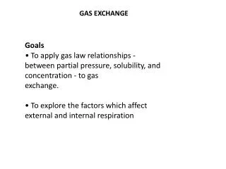
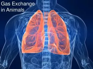
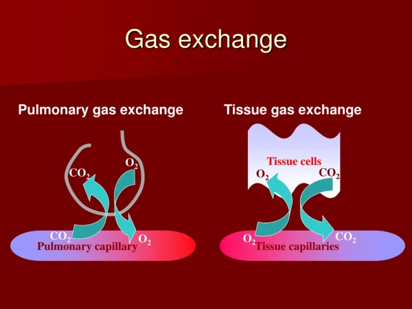
Gas Exchange in Mammals • Delivery of O2 to gas exchange surface (alveoli) and removal of CO2 from capillaries and out of the body (ventilation).
In Larger Organisms • Diffusion alone cannot meet the needs of the inner-most cells of larger organisms. They have a higher demand for O2 and removal of CO2. • Larger organisms must have some form of internal transport system for gases.
Pathway Air (outside) nasal passages (or mouth) pharynx larynx trachea bronchus bronchiole alveoli bloodstream
Nasal passages - Air is warmed and moistened. Dust and bacteria removed by mucous and nasal hairs (cilia).
Cilia - creates wave-like movement to sweep mucous upwards to be expeled at the mouth/nose (along with trapped debris)
Cilia animation • http://www.bioscope.org/taste/cd1/a0255a.htm
Hollow tube held open by ‘C’ shaped bands of cartilage. It branches into the two bronchi (one: bronchus) which also have cartilaginous rings. Trachea
Alveolus Bronchus then branch into smaller tubes called bronchioles which lead into grape-like clusters of thin-walled air sacs called alveoli (one: alveolus) which are surrounded by blood capillaries.
Oxygen diffuses across the moist lining of the alveoli into the capillaries. • CO2 diffuses out of the capillary and into the air in the alveoli.
Surfactant • Lipoprotein produced by alveolar cells • Reduced surface tension (if no surfactant – lung cannot inflate normally)
Oxygen is picked up by a red pigment called haemogoblin (in red blood cells) and is carried around the body in the circulatory system . As the blood carrying the oxygen (oxygenated blood) passes through the body the haemoglobin releases oxygen which diffuses into the cells. Pigment
Other bits • Diaphragm is a sheet of muscle at bottom of these cavities • Pleural membrane lines each cavity and covers each lung, and encloses pleural space (which contains fluid to help membranes slide past each other during breathing) www.tcnj.edu/~mckinney
Inhalation – Breathing In • Diaphragm contracts (lowered), intercostal muscles contract - lifts ribs up and out. • Increases volume of chest cavity cause the air from outside to rush in and fill up the increased space.
Exhalation – Breathing out • Diaphragm and intercostal muscles relaxes • Diaphragm moves up and the ribs down • Air is forced out as the volume in the chest cavity returns to normal.
The breathing action (diaphragm etc.) uses considerable energy but allows the animal to change the rate of gas exchange quickly to suit activity/environment requirements.
Increased Surface Area for Gas Exchange • Alveoli ↑’s surface area exposed for diffusion of O2 into capillaries and CO2 out into alveoli. • 300 million alveoli creates 40x the surface area of the body.
Moist Surfaces • Moist surface within nasal passages, alveoli and capillaries help O2 in air to dissolve into the watery substance for diffusion into capillaries.
Thin Exchange Surface • Thin surface of alveoli and capillaries ↓’s barrier for diffusion of O2 into capillaries and CO2 out.
A more specialised system is required for the increased size of the animal to deal with: → the inefficiency of diffusion over longer distances → higher demand for O2 and removal of CO2. Internal lungs are well protected from: → physical damage → drying out in a dry environment. Enables mammals to have a wider range of habitats e.g. dry, wet, water on land. Lungs have a higher surface area to maximise rate of diffusion of O2 into capillaries and removal of CO2. Then the O2 and CO2 is transported around the body in the bloodstream of the circulation system.
Some more interesting info/pictures Warning – some pictures are a bit gory – so stop here if you are sensitive to blood and gore.
Cross-section of the lung and heart Figure 23–8
Cilia can be immobilized by smoking Smoker’s lung tissue Normal lung tissue www.orangeusd.k12.ca.us/date/
Cystic fibrosis Faulty chloride channel leads to thick mucus difficult to clear blockage and infection Normal lung tissue Lung tissue from cystic fibrosis patient www.orangeusd.k12.ca.us/date/ www.pathguy.com/lectures
Black lung disease An electron micrograph scan of coal dust (marked by dark patches) in lung tissue infected with black lung disease. A disease found primarily in older coal workers, black lung is characterized by thickening and scarring of lung tissue.
Pneumothorax: air trapped in the chest cavity. Pneumothorax: air trapped in the chest cavity. (Tension pneumothorax: life-threatening) (Tension pneumothorax: life-threatening)