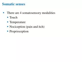
Somatic
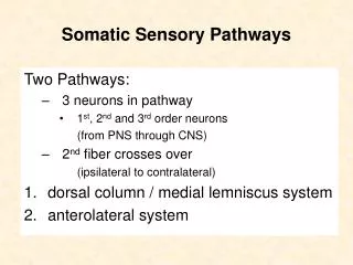
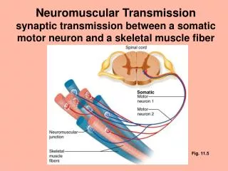
Neuromuscular Transmission synaptic transmission between a somatic motor neuron and a skeletal muscle fiber. Somatic. Fig. 11.5. Neuromuscular Junction. The neuromuscular junction is a chemical synapse at which a nerve impulse triggers the excitation of skeletal muscle.


Somatic
E N D
Presentation Transcript
Neuromuscular Transmissionsynaptic transmission between a somatic motor neuron and a skeletal muscle fiber Somatic Fig. 11.5
Neuromuscular Junction • The neuromuscular junction is a chemical synapse at which a nerve impulse triggers the excitation of skeletal muscle. • motor neuron = presynaptic cell • at the motor neuron: • electrical signal chemical signal • skeletal muscle fiber = postsynaptic cell • at the skeletal muscle fiber: • chemical signal electrical signal
Neuromuscular JunctionThe anatomical structure of the neuromuscular junction is called the motor end plate. Fig. 11.6
Neuromuscular Transmission At the neuromuscular junction, the neurotransmitter released from the motor neuron is acetylcholine (ACh). &The ACh is received by a nicotinic cholinergic receptor. The nicotinic receptor is a cation channel, that allows Na+ to enter the cell; Na+ entry causes depolarization. Fig. 11.7
The Nicotinic Cholinergic Receptor Na+ and Ca++ ACh Fig. 15-15, Alberts et al., Molecular Biology of the Cell Fig. 4-18 Ganong
Neuromuscular Transmission (cont’d) This depolarization, called an end plate potential, is an example of an excitatory postsynaptic potential (EPSP - see graded potentials later). If the EPSP causes the area next to the motor end plate to depolarize to threshold, an action potential is generated. Fig. 11.7
Fig. 11.8 Excitation – Contraction Coupling - DHP receptor The muscle action potential activates the T-tubules’ voltage sensors, the dihydropyridine (DHP) receptors. • DHP is a prototypical calcium channel blocker. • In cardiac and smooth muscle, the DHP receptor is a functional voltage-gated Ca++ channel. • However, in skeletal muscle, the DHP receptor does not function as a Ca++ channel. It is only a voltage sensor.
Fig. 11.8 Excitation – Contraction Coupling - DHP receptor The activated DHP receptors cause the calcium release channels (ryanodine receptors) of the SR to open. • direct coupling? • the favored model for skeletal muscle • via a second messenger (e.g., calcium-induced calcium release)? • the favored model for cardiac muscle Calcium enters the cytosol. ryanodine receptors (not shown)
Triad Structures Direct-coupling Model ryanodine receptors: terminal cistern of SR (SR) DHP receptor ryanodine receptor Alberts, et al, Molecular Biology of the Cell (cf. Fig. 3-8 Ganong)
Fig. 11.8 Excitation – Contraction Coupling - DHP receptor Calcium binds to troponin, and allows actin and myosin to interact. Filaments slide; the muscle contracts. ryanodine receptors
End of Excitation The ACh lasts only a short time because it is broken down by an acetylcholinesterase. The ACh-esterase is anchored to the postsynaptic membrane by a glycolipid. Fig. 11.10
Protein Anchored to Membrane via a Glycolipid Fig. 10-17, Alberts et al., Molecular Biology of the Cell