Download
1 / 70
700 likes | 902 Views
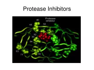
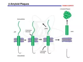
The aspartyl protease BACE b -Amyloid cleaving enzyme. BACE is expressed mostly in the brain. Vassar et al., 1999. In the cell, BACE localizes to Golgi apparatus and Endosomes. Vassar et al., 1999. BACE activity. 1- In vitro, BACE is mostly active at an acidic pH range between 4.5-5.5.

E N D
The aspartyl protease BACE b-Amyloid cleaving enzyme
BACE is expressed mostly in the brain Vassar et al., 1999
In the cell, BACE localizes to Golgi apparatus and Endosomes Vassar et al., 1999
BACE activity 1-In vitro, BACE is mostly active at an acidic pH range between 4.5-5.5. 2-BACE is supposed to be mostly active in the endosomes, due to BACE co-localization and to the acidic pH of these organelles. Although in vivo, interaction between BACE and APP was observed at the plasma membrane and in the endosomes, in cell culture, BACE was active also in the ER and in the Golgi apparatus.
BACE Domains and trafficking 1 aa460-476 501 Propeptide sequence DTG DSG TM furin DDISLLK Regulation of BACE Trafficking Abeta?
The LL motif, but not the S (that can be phoshorylated) regulates the amount of BACE retained at the plasma membrane….. Pastorino et al., MCN 2002
BACE LL motif determines lysosomal colocalization for degradation Koh et al., 2005
GGA proteins: a crucial role in the regulation of BACE trafficking and degradation through BACE LL domain
Do GGA3 and BACE levels change during neurodegenerative pathologies?
Ischemic patients have increased levels of BACE in the brain… Tesco et al., Neuron. 2007 Jun 7;54(5):721-37.
…and decreased levels of GGA3 Tesco et al., Neuron. 2007 Jun 7;54(5):721-37.
AD patients have increased levels of BACE and decreased levels of GGA3 in the brain Tesco et al., Neuron. 2007 Jun 7;54(5):721-37.
GGA3 siRNA causes increase of BACE expression and accumulation of C99 Tesco et al., Neuron. 2007 Jun 7;54(5):721-37.
APP contains caspase cleavage sites in its sequence Tesco et al., Neuron. 2007 Jun 7;54(5):721-37. HOWEVER Although apoptosis increases C99 and Ab levels, this effects do not depend on caspase-mediated cleavage of APP (Tesco et al., 2003).
Apoptosis increases levels of C99….. Tesco et al., Neuron. 2007 Jun 7;54(5):721-37.
…and BACE Tesco et al., Neuron. 2007 Jun 7;54(5):721-37.
During apoptosis GGA3 levels are destabilized Tesco et al., Neuron. 2007 Jun 7;54(5):721-37.
Apoptotic mechanisms associated with neurodegeneration stabilize BACE via the inhibition of GGA3, therefore inhibiting GGA3-mediated BACE degradation
Model of BACE stabilization during apoptosis Vassar, Neuron. 2007 Jun 7;54(5):671-3. Review.
g-secretase complex and the phenomenon of INTRAMEMBRANE PROTEOLYSIS
The g-secretase complex -PS1 and PS2: 7- to 8- transmebrane domain proteins, are the catalytic unit of the g-secretase complex (they will cleave the substrates). -Nicastrin: a type 1a transmembrane protein, approximately 130kDa molecular weight, binds to active PS1, and can sort to the plasma membrane with PS1. Nicastrin has a large extracellular domain crucial for the identification of the substrate. -APH1: (antherior pharinx defective phenotype). Its knockout leads to developmental deficit in C.elegans. It is required for the correct localization of the mature form of nicastrin at the cell surface. Pen2: (presenilin enhancer 2). Stabilizes mature presenilin and nicastrin. The gene knockout for one of any protein of the complex will result in lack of g-secretase activity.
The members of the g-secretase complex Bart Der Strooper
Evidences that the only presenilin is not enough to generate g-secretase activity 1-Overexpression of PS1 in cells does not increase g-secretase dependent cleavage of APP. 2-In yeast, a system known to have NO ENDOGENOUS g-secretase related activity, processing of APP can be observed ONLY when all the four protein of the complex are exogenously expressed.
Model for intramembrane proteolysis Christian Haass
Presenilin: an active heterodimer in the g-secretase complex The Regulated Intramembrane Proteolysis Loop domain Loop domain Endoproteolysis H2O, substrates Modified from Michael Wolfe
g-secretase cleaves different substrates Bart De Strooper
g-secretase dependent intramembrane proteolysis on multiple substrates Xia and Wolfe
Notch is a protein regulating transcription of proteins crucially involved during early and late stages of development Selkoe and Kopan
Like APP, Notch is processed via a- and g-secretase, while trafficking within the cell At the plasma membrane a-secretase After endocytosis g-secretase Selkoe and Kopan
Roles of PS1 in the cell: a switch between physiological and pathological functions in AD Catalytic subunit of the g-secretase: in AD gain of toxic function APP: generation of beta amyloid peptides in AD. NOTCH: production of NICD and control on protein transcription, might be disrupted in AD. Cadherins: control over signaling for cell growth, might be disrupted in AD. Erb4: EGF signaling, might be disrupted in AD. Other mechanisms of toxicity by loss of physiological function?
Autophagic Vacuoles (AVs) accumulate in AD A defective fusion of AVs to the membrane of the lysosomes caused by a loss of function of PS1?
PS1 deletion selectively inhibits macrophagic turnover of proteins Short lived proteins (A-D) PS1 KO causes Inhibition of proteolysis that cannot be further reduced by treatment with 3MA PS1 is involved in the autophagic-dependent proteolysis of substrates. Long lived proteins 3MA blocks the formation of AVs, thus selectively inhibits macroautophagy NH4Cl neutralizes lysosomal pH, thus blocks lysosomal proteolysis
PS1 deletion is insensitive to macroautophagy stimuli (F,G) PS1 KO increases the number of AVs, without affecting the pathways that lead to formation of the autophagosomes (H) Serum withdrawal induces autophagy
Non-mature autophagosomes or autolysosomes accumulate in PS1KO blastocysts Engulfed material in autolysosomes from PS1KO }
Defective autophagy in PS1KO is due to improper fusion of the AVs to the lysosomes
Impairment of PS1 activity in PS1KO cells results in defective acidification of the lysosomes (increased pH), impairing the autophagosomes to fuse with the lysosomes and to allow degradation of the phagosomes’ content.
What’s the implication in AD? Evidences for improper lysosomal proteolysis in PS1 FAD A possible loss of physiological function of PS1 in AD
Autophagy could be protective from AD, PS1 loss of physiological function may be involved in AD pathology
Abeta-42 induces formation of large autophagic vacuoles wt Ab42