Download
1 / 23
240 likes | 525 Views
Amyloid fibrils. Aldo Rampioni Meeting EMBIO project, Vienna, May 2006. What is the signature of amyloid fibrils?. Fibrils bind the dye Congo red, resulting in a green birefringence under polirized light

E N D
Amyloid fibrils Aldo Rampioni Meeting EMBIO project, Vienna, May 2006
What is the signature of amyloid fibrils? • Fibrils bind the dye Congo red, resulting in a green birefringence under polirized light • Electron Microscopy and Atomic Force Microscopy reveal that fibrils are typically long, unbranched and ≈ 100 Å in diameter • X-ray diffraction indicates an ordered cross-β structure which consists of β-sheets running parallel (and the peptides strands perpendicular) to the fibril axis
From cryo-EM: Ribbon-like structure whose height is always multiple of ≈ 21 Å multiple layers From X-ray diffraction: A characteristic distance of ≈ 5 Å between β-strands in the same β-sheet Two distances around 1 nm that arise from face-to-face separation of β-sheets Image from M.L. de la Paz et al. Proc. Natl. Acad. Sci. USA, vol 99, 2002
OPEN PROBLEMS: • STRUCTURE AT ATOMIC DETAIL • PROCESS OF FORMATION • TOXICITY
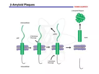
Three-dimensional structure of the transthyretin-retinol-binding protein complex. The tetrameric transthyretin is shown in blue, light blue, green and yellow. The retinol binding protein is shown in red and the retinol is shown in grey. TTR is found in plasma and cerebrospinal fluid and is composed of four identical 127 aminoacid subunits. Function: the tetrameric structure binds and transports thyrosine and retinol binding protein.
To develop a model of fibril I am performing the following simulations in explicit solvent: • Improper Dihedral Restraints on one peptide (in order to • let it assume the structure determined by using NMR) • Distance Restrains among peptides (in order to bring them • close, but stillfree to arrange) The main idea is to restraint the system in order to get an amyloid-like structure (trying both parallel and antiparallel beta sheets) letting it free to arrange… …Until we get a stable multi-layer fibril and we can remove the restraints
From NMR we have informations about the structure of the single peptide in the fibril: Improper dihedrals (choosen such that the experimental error on dihedral angles is of the order of kT, i.e. stochatstic perturbation) Image from C.P. Jaroniec et al. Proc. Natl. Acad. Sci. USA, vol 101, p.711, 2004
We introduce distance restraints compatible with X-ray diffraction data:
So far we have performed short simulations (5 or 10 ns) for systems composed by parallel 2,3,4,5,6,12,24 peptides antiparallel • On which we have changed restraints: • applying improper dihedrals on all peptides or just one • changing the window and the multiplicative constant of distance restraints
4 antiparallel peptides K=0.2 6 antiparallel peptides K=1
5 parallel peptides k=0.2 4 parallel peptides k=2 6 parallel peptides k=1
4 layers of 6 antiparallel peptides
Is it possible to simulate the b-sheet aggregate? Molecular Dynamics simulations to investigate stability of suprastructures of SIVwt peptide aggregates in DMSO Simulation box with 30 parallel chains of SIVwt peptide in DMSO. Amount of particles: ~ 30000 Frame at Tinitial Distance between chain #1 and #30: ~12 nm The simulations show the spontaneous helical twisting of the protofibril Patrica Soto
Spontaneous twisting… t = 0 ns t = 10 ns t = 20 ns t = 35 ns • Parallel or antiparallel? • Chirality • Twisted ribbon as energetically more favorable geometry t = 50 ns Patrica Soto
Is it possible to simulate the spontaneous aggregation into ordered b-sheet aggregates? Solvent: hexane t = 20ns t = 50ns t = 0ns t = 2 ns t = 75ns t = 90ns SIVwt peptide spontaneously aggregates into stable clusters that exhibit high population of b-sheet secondary structure Patrica Soto
Is it possible to simulate the spontaneous aggregation into ordered b-sheet aggregates? Solvent: water t = 2ns t = 0ns t = 35ns t = 50ns SIVwt peptide spontaneously aggregates into stable clusters that exhibit high population of b-sheet secondary structure Patrica Soto
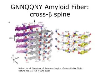
System studied: 7-residue peptide from Sup35 Gly Asn Asn Gln Gln Asn Tyr Picture from Nelson R. et al., 2005, Nature, 435, 773-778
This project is in collaboration with: • Xavier Periole, University of Groningen, The Netherlands • Alan Mark, University of Queensland, Brisbane, Ausralia • Michele Vendruscolo, University of Cambridge, UK
Image from H.D. Nguyen and C.K. Hall . Proc. Natl. Acad. Sci. USA, vol 101, 2004