Pituitary disorder
pituitary insufficiency Dr.Aishah AlEkhzaimy , MBBS,FRCPC,FACE Assistant Professor, Endocrinology consultant. Pituitary disorder. Non-functional pituitary tumor mass-effect Prolactin secreting cell disorder: prolactinoma Growth hormone secreting cell disorder: acromegaly

Pituitary disorder
E N D
Presentation Transcript
pituitary insufficiencyDr.AishahAlEkhzaimy, MBBS,FRCPC,FACEAssistant Professor, Endocrinology consultant
Pituitary disorder • Non-functional pituitary tumor mass-effect • Prolactin secreting cell disorder: prolactinoma • Growth hormone secreting cell disorder: acromegaly • ACTH secreting cell disorders: cushing’s • TSH secreting cell tumor: TSHoma • Gonadotropin secreting cell disorder • Diabetes Insipidus
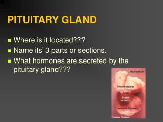
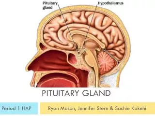
Pituitary Development • Anterior pituitary is recognizable by 4- 5th wk of gestation • Full maturation by 20th wk • From Rathke’s pouch, Ectodermalevagination of oropharynx • Migrate to join neurohypophysis • Portion of Rathke’s pouch →→ Intermediate lobe • Remnant of Rathke’s pouch cell in oral cavity →→ pharyngeal pituitary • Lies at the base of the skull as sellaturcica • Roof is formed by diaphragmasellae • Floor by the roof of sphenoid sinus
Pituitary Development • Posterior pituitary from neural cells as an outpouching from the floor of 3rd ventricle • Pituitary stalk in midline joins the pituitary gland with hypothalamus that is below 3rd ventricle • Development of pituitary cells is controlled by a set of transcription growth factors like pit-1, Prop-1, Pitx2
Pituitary Development • Pituitary stalk and its blood vessels pass through the diaphragm • Lateral wall by cavernous sinus containing III, IV, VI, V1, V2 cranial nerves and internal carotid artery with sympathetic fibers. Both adjacent to temporal lobes • Pituitary gland measures 15 X 10 X 6 mm, weighs 500 mg but about 1 g in women • Optic chiasm lies 10 mm above the gland and anterior to the stalk • Blood supply : superior, middle, inferior hypophysial arteries ( internal carotid artery) running in median eminence from hypothalamus • Venous drainage: to superior and inferior petrosalsinsuses to jugular vein
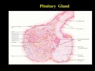
Normal Pituitary Anatomy Modified from Lechan RM. Neuroendocrinology of Pituitary Hormone Regulation. Endocrinology and Metabolism Clinics 16:475-501, 1987
Anterior Pituitary Function Adapted from: William’s Textbook of Endocrinology, 10th ed., Figure 8-4, pg 180.
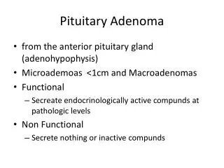
Etiology of Pituitary-Hypothalamic Lesions • Non-Functioning Pituitary Adenomas • Endocrine active pituitary adenomas • Prolactinoma • Somatotropinoma • Corticotropinoma • Thyrotropinoma • Other mixed endocrine active adenomas • Malignant pituitary tumors: Functional and non-functional pituitary carcinoma • Metastases in the pituitary (breast, lung, stomach, kidney) • Pituitary cysts: Rathke's cleft cyst, Mucocoeles, Others • Empty sellasyndrome • Pituitary abscess • Lymphocytic hypophysitis • Carotid aneursym \
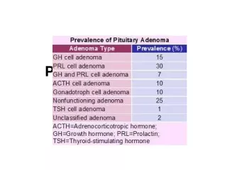
Evaluation of Pituitary mass • Pituitary adenoma: 10 % of all pituitary lesions • Genetic-related • MEN-1, Gs-alpha mutation, PTTG gene, FGF receptor-4 • Pituitary incidentaloma: 1.5 -31% in autopsy ( prevalence) • 10 % by MRI most of them < 1 cm
Evaluation of Pituitary lesion • Functional adenoma ( hormonal-secreting) • Non-Functional adenoma
Evaluation of Pituitary lesion • Non-Functional pituitary lesion: • Absence of signs and symptoms of hormonal hypersecretion • 25 % of pituitary tumor • Needs evaluation either micro or macroadenoma • Average age 50 – 55 yrs old, more in male
Non- functional pituitary adenoma • Presentation of NFPA: • As incidentaloma by imaging • Symptoms of mass effects ( mechanical pressure) • Hypopituitarism ( mechanism) • Gonadalhypersecretion
Non- functional pituitary adenoma • Treatment: • Surgery if indicated - recurrence rate 17 % if gross removal, 40 % with residual tumor - predictors of recurrence: young male, cavernous sinus invasion, extent of suprasellarextention of residual tumor, duration of follow up, marker; Ki-67 • Observation with annual follow up for 5 years and then as needed, visual field exam Q 6-12 month if close to optic chiasm. Slow growing tumour • Adjunctive therapy: - Radiation therapy - Dopamine agonist - Somatostatin analogue
Prolactinoma • Prolactin:
Prolactinoma • Diagnosis: High prolactin after excluding other causes • Management: Dopamine agonist Surgery if no response Radiation therapy
Growth hormone • Pituitary tumor as mass effect →→ Growth hormone deficiency • Hyperfunctioning mass →→ Acromegaly
Growth hormone deficiency • Diagnosis in children and adult
Diagnosis of GH-deficiency and management • GH, IGF-I level • Dynamic testing: clonidine stimulation test, glucagon stimulation, exercise testing, arginine-GHRH, insulin tolerance testing • X-ray of hands: delayed bone age • In Adult: Insulin tolerance testing, MRI pituitary to rule out pituitary adenoma • Management: GH replacement
Acromegaly • Clinical picture and presentation • GH level ( not-reliable, pulsatile) • IGF-I • 75 g OGTT tolerance test for GH suppression • Fasting and random blood sugar, HbA1c • Lipid profile • Cardiac disease is a major cause of morbidity and mortality • 50 % died before age of 50 • HTN in 40% • LVH in 50% • Diastolic dysfunction as an early sign of cardiomyopathy
Growth hormone disorder-Acromegaly • Medical treatment: • Somatostatin analogue • Surgical resection of the tumor
HPA-axis • 2nd adrenal insufficiency • glucgocorticoid replacement • Circadian rhythm of cortisol secretion • Early morning cortisol between 8-9 am
Hypoadrenalism • Nausea • Vomiting • Abdominal pain • Diarrhoea • Muscle ache • Dizziness and weakness • Tiredness • Weight loss • Hypotension
Hypoadrenalism • Diagnosis: Low ACTH and Low morning cortisol • Stimulation test: Insulin tolerance test • Management: Steroid replacement
HPA-axis ( excessive cortisol) • 80 % HTN • LVH • Diastolic dysfunction, intraventricularseptal hypertrophy • ECG needed: high QRS voltage, inverted T-wave • Echocardiogram preop • OSA: 33% mild, 18% severe. Needs respiratory assessment and careful use of sedative during surgery • Glucose intolerance in 60%, control of hyperglycemia • Osteoporosis with vertebral fracture→→ positioning of patient in OR ( 50 %), 20 % with fracture • thin skin→→ difficult IV cannulation, poor wound healing