Download
1 / 110
1.12k likes | 1.18k Views
Explore the autonomic nervous system's anatomy, functions, and reflex actions on target organs, including sympathetic and parasympathetic divisions. Understand neurotransmitters, receptors, and dual innervation. Discover how biofeedback techniques help control responses and maintain homeostasis.

E N D
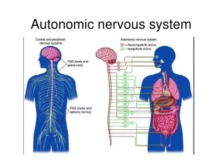
Autonomic Nervous System and Visceral Reflexes • Autonomic nervous system (ANS) • general properties • anatomy • Autonomic effects on target organs • Central control of autonomic function
ANS - General Properties • Motor nervous system controls glands, cardiac and smooth muscle • also called visceral motor system • Regulates unconscious processes that maintain homeostasis • BP, body temperature, respiratory airflow • ANS actions are automatic • biofeedback techniques • train people to control hypertension, stress and migraine headaches
Visceral Reflexes • Unconscious, automatic responses to stimulation of glands, cardiac or smooth muscle • Receptors • detect internal stimuli -- stretch, blood chemicals, etc. • Afferent neurons • connect to interneurons in the CNS • Efferent neurons • carry motor signals to effectors • ANS is the efferent neurons of these reflex arcs • Effectors • glands, smooth or cardiac muscle • ANS modifies effector activity
Divisions of ANS • Two divisions innervate same target organs • may have cooperative or contrasting effects • Sympathetic division • prepares body for physical activity • increases heart rate, BP, airflow, blood glucose levels, etc • Parasympathetic division • calms many body functions and assists in bodily maintenance • digestion and waste elimination • Autonomic tone is the normal rate of activity that represents the balance of the two systems • Effects of each depend upon neurotransmitters released
Sympathetic Nervous System • Origin of presynaptic neurons • lateral horns of spinal cord (T1-L2) • Sympathetic chain ganglia (paravertebral) • 3 cervical, 11 thoracic, 4 lumbar, 4 sacral and 1 coccygeal ganglia • white and gray communicating rami suspend ganglia from spinal nerve • pathways of preganglionic fibers • enter ganglia and synapse on postganglionic cell • travel to higher or lower ganglia and synapse • pass through chain without synapsing to reach collateral ganglia via splanchnic nerves
Sympathetic Nervous System • Neuronal divergence predominates • each preganglionic cell branches and synapses on multiple postganglionic cells • produces widespread effects on multiple organs
Parasympathetic Nervous System • Origin of preganglionic fibers • pons and medulla (for cranial nerve nuclei) • sacral spinal cord segments S2-S4 • Pathways of preganglionic fibers • cranial nerves III, VII, IX and X • arising from sacral spinal cord • pelvic splanchnic nerves and inferior hypogastric plexus • Terminal ganglia in/near target organs • long preganglionic, short postganglionic fibers
Parasympathetic Cranial Nerves • Oculomotor nerve (III) • narrows pupil and focuses lens • Facial nerve (VII) • tear, nasal and salivary glands • Glossopharyngeal (IX) • parotid salivary gland • Vagus nerve (X) • viscera as far as proximal half of colon • Cardiac, pulmonary, and esophageal plexus
Neurotransmitters and Receptors • Effects of ANS • determined by types of neurotransmitters released and types of receptors on target cells • Sympathetic has longer lasting effects • neurotransmitters persist in synapse and some reach the bloodstream • Many substances released as neurotransmitters • enkephalin, substance P, neuropeptide Y, neurotensin, nitric oxide (NO) • NO inhibits muscle tone in BV walls (vasodilation)
Cholinergic Receptors for ACh • Acetylcholine (Ach) binds to 2 classes of receptors • nicotinic receptors • on all ANS postganglionic neurons, in the adrenal medulla, and at neuromuscular junctions (skeletal muscle) • excitatory when ACh binding occurs • muscarinic receptors • on all gland, smooth muscle and cardiac muscle cells that receives cholinergic innervation • excitatory or inhibitory due to subclasses of muscarinic receptors
Adrenergic Receptors for NE • Norepinephrine binds to 2 classes of receptors • alpha adrenergic receptors (often excitatory) • beta adrenergic receptors (often inhibitory) • Exceptions • existence of subclasses of each receptor type • alpha 1 and 2; beta 1 and 2 • Function by means of 2nd messengers • cyclic AMP and alpha 1 receptors
Dual Innervation • Most of viscera receive nerve fibers from both parasympathetic and sympathetic divisions • Both divisions do not normally innervate an organ equally
Dual Innervation • Antagonistic effects • oppose each other • exerted through dual innervation of same effector • heart rate decreases (parasympathetic) • heart rate increases (sympathetic) • exerted because each division innervates different cells • pupillary dilator muscle (sympathetic) dilates pupil • constrictor pupillae (parasympathetic) constricts pupil
Dual Innervation • Cooperative effects seen when 2 divisions act on different effectors to produce a unified effect • parasympathetics increase salivary serous cell secretion • sympathetics increase salivary mucous cell secretion
Control of Autonomic Function • ANS regulated by several levels of CNS • cerebral cortex has an influence • hypothalamus (major visceral motor control center) • nuclei for primitive functions – hunger, thirst • midbrain, pons, and medulla oblongata • nuclei for cardiac and vasomotor control, salivation, swallowing, sweating, bladder control, and pupillary changes • spinal cord reflexes • defecation and micturition reflexes integrated in cord • brain can inhibit these responses consciously
Sense Organs • Sensory receptors • properties and types • General senses • Chemical senses • Hearing and equilibrium • Vision
Properties of Receptors • Sensory transduction • convert stimulus energy into nerve energy • Receptor potential • local electrical change in receptor cell • Adaptation • conscious sensation declines with continued stimulation
Receptors Transmit Information • Modality - type of stimulus • Location • each sensory receptor receives input from its receptive field • sensory projection - brain identifies site of stimulation • Intensity • frequency, number of fibers and which fibers • Duration - change in firing frequency over time • phasic receptor - burst of activity and quickly adapt (smell and hair receptors) • tonic receptor - adapt slowly, generate impulses continually (proprioceptor)
Classification of Receptors • By modality: • chemo-, thermo-, mechano-, photo- receptors and nociceptors • By origin of stimuli • interoceptors - detect internal stimuli • proprioceptors - sense body position and movements • exteroceptors - detect external stimuli • By distribution • general senses - widely distributed • special senses - limited to head
Unencapsulated Nerve Endings • Dendrites not wrapped in connective tissue • General sense receptors • for pain and temperature • Tactile discs • associated with cells at base of epidermis • Hair receptors • monitor movement of hair
Encapsulated Nerve Endings • Dendrites wrapped by glial cells or connective tissue • tactile corpuscles - phasic • light touch and texture • krause end bulb - phasic • tactile; in mucous membranes • lamellated corpuscles - phasic • deep pressure, stretch, tickle and vibration • ruffini corpuscles - tonic • heavy touch, pressure, joint movements and skin stretching
Somesthetic Projection Pathways • 1st order neuron (afferent neuron) • from body, enter the dorsal horn of spinal cord via spinal nerves • from head, enter pons and medulla via cranial nerve • touch, pressure and proprioception on large, fast, myelinated axons • heat and cold on small, unmyelinated, slow fibers • 2nd order neuron • decussation to opposite side in spinal cord or medulla/pons • end in thalamus, except for proprioception (cerebellum) • 3rd order neuron • thalamus to primary somesthetic cortex of cerebrum
Pain • Nociceptors – allow awareness of tissue injuries • found in all tissues except the brain • Fast pain travels in myelinated fibers at 30 m/sec • sharp, localized, stabbing pain perceived with injury • Slow pain travels unmyelinated fibers at 2 m/sec • longer-lasting, dull, diffuse feeling • Somatic pain from skin, muscles and joints • Visceral pain from stretch, chemical irritants or ischemia of viscera (poorly localized) • Injured tissues release chemicals that stimulate pain fibers (bradykinin, histamine, prostaglandin)
Projection Pathway for Pain • General pathway – conscious pain • 1st order neuron cell bodies in dorsal root ganglion of spinal nerves or cranial nerves V, VII, IX, and X • 2nd order neurons decussate and send fibers up spinothalamic tract or through medulla to thalamus • gracile fasciculus carries visceral pain signals • 3rd order neurons from thalamus reach primary somesthetic cortex as sensory homunculus • Spinoreticular tract • pain signals reach reticular formation, hypothalamus and limbic • trigger visceral, emotional, and behavioral reactions
Referred Pain • Misinterpreted pain • brain “assumes” visceral pain is coming from skin • heart pain felt in shoulder or arm because both send pain input to spinal cord segments T1 to T5
CNS Modulation of Pain • Intensity of pain - affected by state of mind • Endogenous opiods (enkephalins, endorphins and dynorphins) • produced by CNS and other organs under stress • in dorsal horn of spinal cord (spinal gating) • act as neuromodulators block transmission of pain
Spinal Gating • Stops pain signals at dorsal horn • descending analgesic fibers from reticular formation travel down reticulospinal tract to dorsal horn • secrete inhibitory substances that block pain fibers from secreting substance P • pain signals never ascend • dorsal horn fibers inhibited by input from mechanoreceptors • rubbing a sore arm reduces pain
Chemical Sense - Taste • Gustation - sensation of taste • results from action of chemicals on taste buds • Lingual papillae • filiform (no taste buds) • important for texture • foliate (no taste buds) • fungiform • at tips and sides of tongue • vallate (circumvallate) • at rear of tongue • contains 1/2 of taste buds
Taste Bud Structure • Taste cells • apical microvilli serve as receptor surface • synapse with sensory nerve fibers at their base • Supporting cells • Basal cells
Physiology of Taste • Molecules must dissolve in saliva • 5 primary sensations - throughout tongue • Sweet - concentrated on tip • Salty - lateral margins • Sour - lateral margins • Bitter - posterior • Umami - taste of amino acids (MSG) • Influenced by food texture, aroma, temperature, and appearance • mouthfeel - detected by lingual nerve in papillae • Hot pepper stimulates free nerve endings (pain)
Physiology of Taste • Mechanisms of action • activate 2nd messenger systems • sugars, alkaloids and glutamates bind to receptors • depolarize cells directly • sodium and acids penetrate cells
Projection Pathways for Taste • Innervation of taste buds • facial nerve (VII) - anterior 2/3’s of tongue • glossopharyngeal nerve (IX) - posterior 1/3 • vagus nerve (X) - palate, pharynx, epiglottis
Chemical Sense - Smell • Olfactory mucosa • contains receptor cells for olfaction • highly sensitive • up to 10,000 odors • on 5cm2 of superior concha and nasal septum
Olfactory Epithelial Cells • Olfactory cells • olfactory hairs neurons with 20 cilia • bind odor molecules in thin layer of mucus • axons pass through cribriform plate • survive 60 days • Supporting cells • Basal cells • divide
Physiology of Smell • Molecules bind to receptor on olfactory hair • hydrophilic - diffuse through mucus • hydrophobic - transport by odorant-binding protein • Activate G protein and cAMP system • Opens ion channels for Na+ or Ca2+ • creates a receptor potential • Action potential travels to brain • Receptors adapt quickly • due to synaptic inhibition in olfactory bulbs
Olfactory Pathway • Olfactory cells synapse in olfactory bulb • on mitral and tufted cell dendrites • in spherical clusters called glomeruli • each glomeruli dedicated to single odor
The Nature of Sound • Sound - audible vibration of molecules • vibrating object pushes air molecules
Pitch and Loudness • Pitch - frequency vibrates specific parts of ear • hearing range is 20 (low pitch) - 20,000 Hz (cycles/sec) • speech is 1500-4000 where hearing is most sensitive • Loudness – amplitude; intensity of sound energy
Outer Ear • Fleshy auricle (pinna) directs air vibrations down external auditory meatus • cartilagenous and bony, S-shaped tunnel ending at eardrum • glandular secretions and dead cells form cerumen (earwax)