Web based tools developed to help students understand cell ultra-structure
230 likes | 363 Views
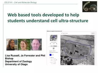
Web based tools developed to help students understand cell ultra-structure. Lisa Russell, Jo Forrester and Phil Bishop Department of Zoology University of Otago. Frog cell at Anaphase. Cell ultra-structure.

Web based tools developed to help students understand cell ultra-structure
E N D
Presentation Transcript
Web based tools developed to help students understand cell ultra-structure Lisa Russell, Jo Forrester and Phil Bishop Department of Zoology University of Otago Frog cell at Anaphase
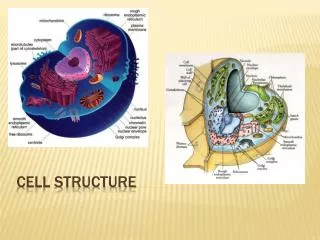
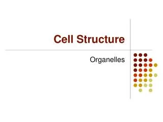
Cell ultra-structure • Develop the students capacity to infer function of different cell types based on their recognition of constituent organelles. Images Katrin Geist and Richard Easingwood
Challenges facing large, first year biology classes: Evident that students struggle to make the link between: • 3-d structure of organs, tissues and cellular components from lectures • 2-d images that they obtain from light and electron micrographs
Challenges facing large, first year biology classes: • Textbook examples of stylised ‘typical’ cells often represent an oversimplification of real life images students will view during the course.
Challenges facing large, first year biology classes: Only a very limited time and resources to provide students with hands-on practical experience with microscopy • limited to light microscopy Viewing electron micrographs (TEM and SEM) restricted: • paper exercises or as web-based activities • freeonline software available has limitations (Image J and Zoomify)
Image J • Open source program • Wide range of tools, including a calibration tool Limitations: • not user friendly • requires detailed step by step instructions to calibrate image, annotate image etc. • students must download program x=1.40, y=0.627, angle=-131.56, length = 5.30
Zoomify • freeware package • high resolution imaging Limitations of package: • unable to calibrate or calculate scale • unable to add annotations or a key
Project Develop a web-based tool to complement guided learning exercises that investigate: • how cell ultra-structure is imaged (microscopy and scale) • structure and function of common organelles • cell diversity and ultra-structural differences (simple versus specialised cells)
Project Collaborative project: • Educational Media, Higher Education Development Centre (Peter Vlugter and Ayelet Cohen) • Otago Centre for Electron Microscopy (OCEM) in particular Mr Richard Easingwood and Miss Katrin Geist Funded by a CALT (Committee for the Advancement of Learning and Teaching) e-learning grant
Project • Development of a range of high resolution EM images from a range of cell types: ‘simple cells’ (plant and animal cells) and specialised cells (secretory and absorptive)
Ideal features of an image viewer: • Zoom and panning tool • Annotation tool • Interactive key • Measuring tool (linear and relative area)
Annotation tool Nucleus Dark stained areas – heterochromatin light areas - euchromatin
Application of the image viewer: • Image viewer is now used in three first year courses Biology 112 (Introduction to animal biology) • Utilized in a practical laboratory • Proportion of red muscle present within a cross section of eel tissue subjected to differing hormonal treatments
Image viewer is now freely available throughout the university as a plug-in to UniTube (file sharing system, wide range of file types including images, videos and audio files). • Once uploaded the UniTube URL can be shared or embed it into Blackboard or any other website.
‘Build a cell’ game • Online interactive game designed to test students ability to link cell structure with function.
‘Build a cell’ game • Four levels - in each level they have to essentially build the specified cell from a range of cell components. • simple plant and animal cell, specialised secretory and absorptive cells.
‘Build a cell’ game To progress through each level: • correct cell components in roughly the correct frequency (1, 2-10 or extensive) • 3 attempts at each level, if incorrect they have to start the game again.
Ongoing developments Image viewer • Develop the annotation tool further • allow students to annotate and save images for reviewing • potential for assessment Build a cell game • developing specific feedback