Download
1 / 54
620 likes | 986 Views
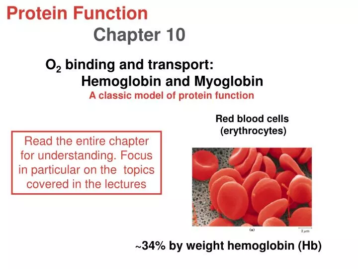
Protein Function Chapter 10. O 2 binding and transport: Hemoglobin and Myoglobin A classic model of protein function. Red blood cells (erythrocytes). Read the entire chapter for understanding. Focus in particular on the topics covered in the lectures.

E N D
Protein Function Chapter 10 O2 binding and transport: Hemoglobin and Myoglobin A classic model of protein function Red blood cells (erythrocytes) Read the entire chapter for understanding. Focus in particular on the topics covered in the lectures ~34% by weight hemoglobin (Hb)
Myoglobin • Small intracellular protein in vertebrate muscle • Oxygen binding protein • Globular protein • First protein whose structure was determined by X-ray crystallography • 153 residues • 8 a helices • Heme is tightly wedged in a hydrophobic pocket between the E and F helices E F
Myoglobin Structure Mb is a monomeric heme protein • Mb polypeptide "cradles" the heme group • Fe in Mb is Fe2+ - ferrous iron - the form that binds oxygen • Oxidation of Fe yields 3+ charge - ferric iron -metmyoglobin does not bind oxygen • Dioxygen binds as the sixth ligand to Fe
Globin -- means without heme Myoglobin’s structure & function provide insights into the structure & function of hemoglobin Hb is a tetramer of 4 Mb-like polypeptides
Heme • is a prosthetic group • ispermanently associated with the protein’s native structure & function • consists of a protoporphyrin ring bound to ferrous iron ( Fe2+) • the iron has six coordination bonds, four to nitrogens within the ring and two perpendicular to the ring • heme is noncovalently bound to protein (Fe-N bond is a coordinate covalent bond)
Porphyrin molecules • Tetrapyrrole macrocyclic compounds • Bind metal ions readily in center, especially iron
Hydrophilicface Propionic acidgroups Vinyl groups Hydrophobic face Iron bound to protoporphyrin IX or “heme”
Fe is in +2 oxidation state whether or not the dioxygen is bound The heme group
O2 N N N N His DeoxyHb His-Fe2+ (color of venous blood) OxyHb His-Fe2+-O2 (color of arterial blood) MetHb His-Fe3+-OH2 (color of old meat) MetHb does not bind dioxygen. Water occupies the 6th coordination site
The visible absorption spectra of oxygenated and deoxygenated hemoglobins Scarlet red arterial blood Dark purple venous blood Page 322 When dioxygen binds, the electronic properties of heme-iron changes, turning fromdark purpletobright red
Dioxygen can only bind to one side of the protein protected heme.
Myoglobin, “Mb” • facilitates dioxygen transport in muscle and NO metabolism • 153 amino acid residues • 8 a-helical segments, A-H • His93 (proximal histidine) binds directly to iron • No other covalent attachments of heme to protein (contrast cytochrome c)
Reversible binding of oxygen to Mb is described by the simple equilibrium reaction PL P + L Protein = P = Mb Ligand = L = O2 Protein-ligand complex = PL Dissociation constant = Kd (= 1/Ka) Kd = [P][L] or [PL] = [P][L] [PL] Kd YO2 = [PL] = [L] [PL]+ [P] [L] + Kd YO2= fractional saturation = fraction of the total binding sites occupied by dioxygen ligand
YO2= [L] = fraction of the total binding [L] + Kdsites occupied by ligand For a gas, the concentration dissolved is proportional to the partial pressure of the gas over the solution. YO2 = pO2 pO2 + P50 P50 = pO2 at half saturation, i.e., [Mb] = [MbO2] This equation represents a rectangular hyperbola
Myoglobin (Mb) oxygen binding curve Mb saturated with oxygen Rectangular hyperbola • Curve shape typical of the simple binding • of a small molecule to a protein • at very low pO2, very little O2 binds to Mb • at very high pO2, Mb is saturated with O2
Besides oxygen, small molecules such as CO, NO & H2S can bind to heme groups in proteins Mb has ~200 times greater affinity for CO than for dioxygen
Myoglobin O2 Binding Site H-bonds to dioxygen Distal (distant) histidine Structures of oxy and deoxy Mb are almost superimposable Proximal (near) histidine Fifth ligand
Hb • Intracellular protein that gives red blood cells their color • Not just a simple O2 tank • Sophisticated delivery system that provides the proper amount of oxygen to tissues under variety of circumstances • Tetramer
The quaternary structure of deoxyhemoglobin From Lehninger Principles of Biochemistry
The 3-D structures of the a and b chains of Hb look like the structure of Mb, but their primary sequences are different.
Tertiary structure of a and b are remarkably similar both to each other and to Mb Only 18% of the corresponding residues are identical among these 3 polypeptides
Hemoglobin (Hb) carries dixygen a chain b chain Hb (a2, b2) Dioxygen binding site The subunit associations are predominantly hydrophobic (although ion pairs and H-bonding are also involved)
P50 of Hb >> Mb Hb – sigmoidal (S-shaped curve) This permits the blood to deliver much more O2 to the tissue than if Hb had a hyperbolic curve with the same P50
In any binding system, a sigmoidal curve is diagnostic of a cooperative interaction between binding sites Binding of one dioxygen molecule increases the affinity of Hb for binding additional dioxygen molecules.
OxyHb DeoxyHb Solvent-filled central channel b chains draw closer together upon oxygenation
Oxygen Binding by Hb A Quaternary Structure Change • When deoxy-Hb crystals are exposed to oxygen, they shatter! Evidence of a structural change! • One alpha-beta pair moves relative to the other by 15 degrees upon oxygen binding • This massive change is induced by movement of Fe by 0.039 nm when oxygen binds
O2 binding alters the structure of entire Hb tetramer • So the structures of deoxyHb and OxyHb are noticeably different • Oxygenation rotates 1 ab dimer about 15o with respect to the other ab dimer • This brings the b subunits closer together
The Conformation Change The secret of Mb and Hb! • Dioxygen binding changes the Mb conformation • Without dioxygen bound, Fe is out of heme plane • Dioxygen binding pulls the Fe into the heme plane • Fe pulls its His F8 ligand along with it • The F helix moves when dioxygen binds • Total movement of Fe is 0.029 nm - 0.29 A • This change means little to Mb, but lots to Hb!
Hb has 2 stable conformational states (T & R) T state– conformation of deoxyHb R state– conformation of oxyHb
Movement of Fe(II) into the heme plane triggers the T to R conformational shift Movements of the heme & the F helix during the T to R transition in Hb T form Fe2+ R form
The trigger for the T to R state conversion Puckered and Fe out of plane Fe moves in plane, pulling proximal His and Helix F Helix F is moved as oxygen binds. This promotes subunit rotation and rearrangementof the a1b2 (and b2a1) interfaces.
Oxy- and deoxy Hb have different quaternary structures Low Affinity T state High Afinity R State • T state (tense) and R state (relaxed) represent two different conformations of the tetramer • Both bind O2 but R state binds it more strongly
Structure of Hb: The a1b1anda2b2interfaces are the strongest 30 residues form the interface between a1b1 (and a2b2). 19 residues form the interface between a1b2 (and a2b1).
Interactions between a1 and 1 and between a2 and 2 are dominant & change little in the T-to-R conformational change. • The major shifts are at the interfaces between a1 and 2 (and a2 and 1)
Ion pairs that stabilize the T state of deoxyhemoglobin must break to form R state
T vs R State (1) 15 degree rotation of a1b1 relative to a2b2 (2) Change at interface between b1a2 and b2a1 (3) R state is more compact (4) T state has additional salt bridges (5) In R state individual O2 sites have higher affinity for O2. - better Fe-O2 bond length - fewer steric repulsions
Hb binds oxygen cooperatively Hb sigmoidal curve is a composite of hyperbolic T and R state curves T state has reduced O2 affinity Due to Fe O2 bond being stretched beyond its normal length by steric repulsions between heme and oxygen Once more than 1 oxygen binds the molecules snaps to R All subunits are snapped to R whether or not all of them are bound with oxygen R State T state
Hill Plots are used to determine degree of cooperativity 4th dioxygen to bind to Hb does so with 100-fold greater affinity than the 1st. Hill constant: normal Hb between 2.8 –3 Abnormal Hb less than 3
The Bohr Effect Competition between oxygen and H+ • Discovered by Christian Bohr • Binding of protons diminishes dioxygen binding • Binding of dioxygen diminishes proton binding • Important physiological significance
Hb is regulated by H+ and CO2 The Bohr effect: the effect of pH and CO2 on the binding and release of oxygen to Hb. Lower pH (higher H+) stabilizes the T-state lungs More O2 released as the pH is lowered tissue
Protonation of His HC3 in T state is a major contributor to Bohr effect
Bohr Effect II Carbon dioxide diminishes dioxygen binding • Hydration of CO2 in tissues and extremities leads to proton production • These protons are taken up by Hb as oxygen dissociates • The reverse occurs in the lungs
CO2 (high in tissues) is also carried by hemoglobin This reaction produces protons and additional salt bridges stabilize the T state.
2,3-BPG and Hb The "inside" story...... • Where does 2,3-BPG bind? • "Inside" • in the central cavity • What is special about 2,3-BPG? • Negative charges interact with 2 Lys, 4 His, 2 N-termini • Fetal Hb - lower affinity for 2,3-BPG, higher affinity for oxygen, so it can get dioxygen from mother
BPG binding stabilizes the T-state of deoxyHb BPG binds in central cavity • BPG decreases dioxygen binding affinity of Hb • Preferentially binds to deoxy state • BPG binding to deoxyHb is ionic in character • BPG shifts the T R equilibrium towards the T state • hence lower affinity for dioxygen
BPG binding stabilizes the T-state of deoxyHb Blue indicates area of high positive charge T-state R-state Binding pocket open Binding pocket closed
BPG in blood normally 5 mM, but it rises at high altitudes An Allosteric Effector of Hemoglobin • In the absence of 2,3-BPG, oxygen binding to Hb follows a rectangular hyperbola! • The sigmoid binding curve is only observed in the presence of 2,3-BPG • Since 2,3-BPG binds at a site distant from the Fe where oxygen binds, it is called an allosteric effector