Download
1 / 18
180 likes | 208 Views
Learn about the anatomy of the ear, from the outer to inner structures, including how hearing and balance function. Discover the pathways of sound and equilibrium in this comprehensive guide.

E N D
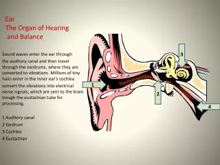
Anatomy of the Ear • The ear is divided into 3 parts: • the outer ear • the middle ear • the inner ear • The outer and middle ear structures are involved with hearing only. • The inner ear functions in both hearing and equilibrium.
Outer (External) Ear • Made up of the pinna and the auditory canal. • The pinna is what most people call the “ear”. The shell-shaped structure surrounds the auditory canal.
The auditory canal is carved into the temporal bone. • In its skin-lined walls are ceruminous glandsthat secrete a waxy, yellow substance called earwax or cerumen. • Sound waves that enter this canal eventually reach the tympanic membrane or eardrum where they cause vibrations. • The eardrum separates the outer ear from the middle ear.
The Middle Ear • It is bordered: • Laterally by the eardrum • Medially by a bony structure with 2 openings* • The structures of the middle ear are: • The Eustachian tube • The ossicles (three small bones) * only one is of importance
Also in the middle ear is the Eustachian tube (AKA: the auditory tube) runs obliquely downward to link the middle ear with the throat. • Typically, this tube is flattened and closed, but yawning or swallowing can temporarily open it up. Doing this will equalize the pressure within the middle ear cavity to that of external environment. • This is important, because when the pressures are unequalled, the eardrum cannot vibrate; this causes hearing difficulty and often earaches.
Ear infections are more common in children than in adults, because their Eustachian tubes are shorter, narrower, and more horizontal than that of adults.
The middle ear also houses the 3 smallest bones (ossicles) in the body. • The ossiclestransmit vibratory motion from the eardrum to the fluid within the inner ear. • These bones are named for their shape: • Hammer or malleus • Anvil or incus • Stirrup or stapes
Transmitting Sound • When the eardrum moves, the hammer moves with it. • The hammer transfers the vibrations to the anvil. • The anvil next passes the vibrations on to the stirrup which presses on the oval window. • Movement at the oval window sets the fluids in the inner ear into motion and this is perceived by nerves called hearing receptors.
The Inner Ear • The inner ear is made up of a combination of parts known as the bony labyrinth. • These bones lie deep within the temporal bone just behind the eye socket. • There are 3 subdivisions to the bony labyrinth: • The cochlea (for hearing) • The vestibule (the connector) • The semicircular canals (for equilibrium)
The special organ of hearing is the cochlea. • The cochlea is a (curled up) tube filled with fluid and lined with hearing receptor cells that pick up the fluid vibration transmitted by the ossicles. • Nerve impulses are created and then transmitted through the vestibularcochlear nerve to the hearing center of the brain.
Pathway of Hearing • Sound waves → pinna → auditory canal → tympanic membrane → ear ossicles→ vibrationsstimulate the receptors in the cochlea → nerve signals travel through (auditory part of ) the vestibulocochlear nerve → temporal lobe of the brain for interpretation.
Also within the inner ear are 3 semicircular canals. • They are also filled with fluid and are lined with nerve cells. • The liquid and neurons identify when there is motion of the head or of the body. • A nerve impulse is then sent to the cerebellum helping to maintain body equilibrium.
Pathway of Equilibrium • Movement of head → stimulates nerve cells in the semicircular canals of the inner ear → nerve signals travel through (vestibular part of) the vestibulocochlear nerve → cerebellum of the brain for interpretation.