Download

1 / 26
260 likes | 342 Views
Explore detailed information on the fascial compartments and muscles of the lower limb, including thigh and leg structures and clinical considerations. Learn about the divisions, contents, nerve supplies, and actions of key muscles. Enhance your anatomical knowledge!

E N D
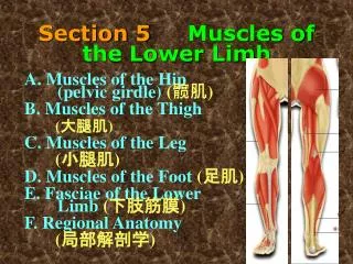
Fascial Compartments of the Thigh Fascia lata is connected to the linea aspera by three intermuscular septa; 1- Medial intermuscular septum 2- Lateral intermuscular septum 3- Posterior intermuscular septum Thus the deep fascia and septa divide the thigh into three compartment; Anterior Posterior Medial
The quadriceps femoris muscle Consisting of: 1- The rectus femoris 2- The vastus intermedius 3- The vastus lateralis 4- The vastus medialis Rectus femoris Vastus lateralis Vastus medialis
The quadriceps femoris muscle Vastus intermedius Remember Quadriceps femoris is the main extensor of the knee joint Nerve supply : femoral nerve Ligamentum patellae
Contents of the medial fascial compartment Muscles ADDUCTOR LONGUS ADDUCTOR BREVIS ADDUCTOR MAGNUS Nerve supply: Obturator nerve Actions:Adducts thigh at hip joint
Contents of the Posterior Fascial Compartment of the Thigh 1-Muscles: Biceps femoris Semitendinosus Semimembranosus 2-Nerve supply: Sciatic nerve Biceps femoris
Semitendinosus Semimembranosus Adductor magnus
Sciatic Nerve • A branch of the sacral plexu • Commonly terminates in the middle of the thigh • by dividing into Tibial Nerve • and Common peroneal
Popliteal Fossa Is a diamond-shaped intermuscular space situated at the back of the knee
Contents of the popliteal fossa • Popliteal artery and vein • The common peroneal nerve • Tibial nerve The popliteal artery AT the middle of the fossa The popliteal artery is the deepest structure While the vein is intermediate and the tibial nerve Is most superficial
Fascial Compartments of the Leg • The deep fascia of the leg divides the leg into Three compartments; • Anterior • Lateral • Posterior • Each having its own muscles, blood supply, and nerve supply.
Contents of the Anterior Fascial Compartment of the Leg Selected Muscles: Extensor digitorum longus Extensor hallucis longus The main actions of these muscles are Extension of the foot at the ankle joint(dorsiflextion) to raise the toes up (in other words to stand up on the heels) Nerve supply of all the muscles of the anterior compartment of the leg: deep peroneal nerve
Contents of the Lateral Fascial Compartment of the Leg • Muscles: • Peroneus longus: • peroneus brevis: • Nerve supply: Superficial peroneal nerve
Contents of the Posterior Fascial Compartment of the Leg • The transverse septa of the legdivides the muscles of the posterior compartment • intosuperficialanddeep groups Deep group of muscles Flexor digitorum longus Flexor hallucis longus Superficial group of muscles Gastrocnemius Soleus Selected muscles Selected muscles Nerve supply: Tibial nerve
Superficial group of muscles • Gastrocnemius • Plantaris • Soleus
Deep group of muscles • Popliteus • Flexor digitorum longus • Flexor hallucis longus • Tibialis posterior
The femoral artery In the femoral triangle, its pulse is easily felt just inferior to the inguinal ligament midway between the pubic symphysis and the anterior superior iliac spine.
The popliteal artery The popliteal artery pulse is difficult to find, but usually can be detected on deep palpation just medial to the midline of the popliteal fossa.
The dorsalis pedis artery Passes onto the dorsal aspect of the foot and anteriorly over the tarsal bones where it lies between and is parallel to the tendon of extensor hallucis longus and the tendon of extensor digitorum longus to the second toe.
The posterior tibial artery Is palpable just posteroinferior to the medial malleolus between the heel and medial malleolus.
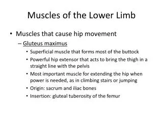
Gluteus medius • Gluteus minimus • Innervation • Superior gluteal nerve
Clinical Notes The great thickness of gluteus maximus muscle makes it ideal for intramuscular injections. To avoid injury to the underlying sciatic nerve, the injection should be given well forward on the upper outer quadrant of the buttock.
However, the upper lateral quadrant, most likely to be made by the Gluteus medius muscle rather than the gluteus maximus muscle . The gluteus maximus covers the posterior part only of the Gluteus medius while the anterior part (which makes the upper lateral quadrant) is covered by skin and fascia only Therefore, the intramuscular injection will be injected into the gluteus medius muscle rather than gluteus maximus muscle