Download
1 / 33
340 likes | 368 Views
Understand the period of isolation for Streptococcal, Pneumococcal, Staphylococcal, and Enterococcal infections recommended by Asst. Professor Dr. P. K. Panda at AIIMS, Rishikesh. Learn about the characteristics, transmission, symptoms, diagnosis, and treatment of Streptococcal Infections. Gain insights into the epidemiology, pathophysiology, clinical syndromes, and differential diagnosis of Pneumococcal Infections. Stay informed on the latest developments in managing these infectious diseases.

E N D
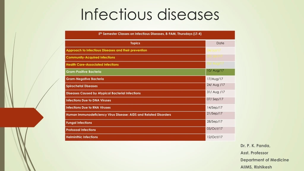
Infectious diseases Dr. P. K. Panda, Asst. Professor Department of Medicine AIIMS, Rishikesh
Streptococcal Infections Pneumococcal Infections Staphylococcal Infections Enterococcal Infections
Streptococcal Infections • Streptococcus, from the Greek streptos, meaning “twisted,” and kokkos, meaning “berry” • Normal flora colonizing the human respiratory, gastrointestinal, and genitourinary tracts • Facultative anaerobes, although some are strict anaerobes, are Fastidious and grow best in 5% CO2 • Respiratory droplets are the usual mechanism of spread, although other routes, including food-borne outbreaks been described
GROUP A STREPTOCOCCI (GAS) • Elaborates a number of cell-surface components (M protein, Polysaccharide capsule) and extracellular products (streptolysins S and O, streptokinase, DNAses,SpyCEP, several pyrogenic exotoxins) • Differential diagnosis of streptococcal pharyngitis includes many, however, Symptoms and signs suggestive of viral infection to be ruled out (conjunctivitis, coryza, cough, hoarseness, or discrete ulcerative lesions of the buccal or pharyngeal mucosa) • The throat culture remains the diagnostic gold standard (properly collected, by vigorous rubbing of a sterile swab over both tonsillar pillars) • A rapid diagnostic kit for latex agglutination or enzyme immunoassay of swab specimens is a useful adjunct with >95% specificity • Treatment is given primarily to prevent suppurativecomplications and ARF AND requires 10 days of penicillin treatment
Complications • Suppurative complications result from the spread of infection from the pharyngeal mucosa to deeper tissues by direct extension or by the hematogenous or lymphatic route and may include cervical lymphadenitis, peritonsillar or retropharyngeal abscess, sinusitis, otitis media, meningitis, bacteremia, endocarditis, and pneumonia • Local complications, such as peritonsillar or parapharyngeal abscess formation, • Nonsuppurativecomplications include ARF and PSGN • ARF is not a sequela to streptococcal skin infections, although PSGN may follow either skin or throat infection. The reason for this difference is not known. • One hypothesis is that the immune response necessary for development of ARF occurs only after infection of the pharyngeal mucosa. • In addition, the strains of GAS that cause pharyngitis are generally of different M protein types than those associated with skin infections. • up to 20% of individuals in certain populations may have asymptomatic pharyngeal colonization • When a carrier is transmitting infection to others, attempts to eradicate carriage are warranted, but no specific treatment/guidelines
Pneumococcal Infections • Belongs to the α-hemolytic group, but growth is inhibited in the presence of optochin (ethylhydrocupreine hydrochloride), and bile soluble • Rapid and dramatic changes in the epidemiology of during the past decade after routine childhood vaccine (PCV) • Not all pneumococcal serotypes are equally likely to cause disease; serotype distribution varies by age, disease syndrome, and geography • Intermittent inhabitants of the healthy human nasopharynx and are transmitted by respiratory droplets • Spread either via the bloodstream to distant sites or locally to mucosal surfaces • In children, nasopharyngeal ecology varies by geographic region, socioeconomic status, climate, degree of crowding, and particularly intensity of exposure to other children
Invasive pneumococcal disease (IPD) • Pathophysiology of severe pneumococcal pneumonia among adults reflects a rapidly progressive cascade of events that often unfolds irrespective of antibiotic administration • Rates of pneumococcal disease vary by season, by sex, and by risk group • Local cytokine production after a viral infection is thought to upregulate adhesion factors • Delayed ontogeny of capsule-specific IgG in young children is associated with susceptibility to infection • There is no pathognomonic presentation of pneumococcal disease • The suspicion should be in differential diagnosis of • pneumonia, • otitis media, • fever of unknown origin, and • meningitis
Clinical syndromes • Classified as noninvasive (e.g., otitis media and nonbacteremic pneumonia) or invasive (e.g., bacteremic pneumonia) • The presentation of pneumococcal pneumonia does not reliably distinguish it from pneumonia of other etiologies • The differential diagnosis includes • Cardiac conditions such as myocardial infarction and heart failure with atypical pulmonary edema; • Pulmonary conditions such as atelectasis; and pneumonia caused by viral pathogens, mycoplasmas, Haemophilusinfluenzae, Klebsiellapneumoniae, Staphylococcus aureus, Legionella, or (in immunocompromised hosts) Pneumocystis • Cholecystitis, appendicitis, perforated peptic ulcer disease, and subphrenicabscesses
Pneumonia • The gold standard for etiologic diagnosis of pneumococcal pneumonia is pathologic examination of lung tissue • Gram’s staining and culture of sputum • Chest radiography helps in diagnosis, but an infiltrate can be absent either early in the course of the illness or with dehydration; upon rehydration, an infiltrate usually appears • The appearance varies; lobar or segmental or patchy consolidation • Parapneumonic effusions are more common than empyema • Pleural fluid with frank pus, bacteria, or a pH of ≤7.1 indicates empyema • Blood cultures are positive in a minority (<30%) • Urinary pneumococcal antigen assays; important among whom nasopharyngeal colonization is relatively low
Meningitis • Pneumococcal meningitis typically presents as a pyogenic condition that is clinically indistinguishable from meningitis of other bacterial etiologies • Definitive diagnosis of pneumococcal meningitis rests on CSF examination • (1) evidence of turbidity (visual inspection); • (2) elevated protein level, elevated white blood cell count, and reduced glucose concentration (quantitative measurement); and • (3) specific identification of the etiologic agent (culture, Gram’s staining, antigen testing, or polymerase chain reaction [PCR]) • Blood culture or detection of antigen in urine is considered highly specific • Mortality rate for pneumococcal meningitis is ~20%
Other Invasive Syndromes • Primary bacteremia without other sites of infection (bacteremia without a source; occult bacteremia), • Osteomyelitis • Septic arthritis • Endocarditis • Pericarditis • Peritonitis • The essential diagnostic approach is collection of fluid from the site of infection by sterile technique and examination by Gram’s staining, culture, and—when relevant—capsular antigen assay or PCR
Noninvasive Syndromes • Otitis media is the most common pneumococcal syndrome and most often affects young children • Sinusitis presents with facial pain, congestion, fever, and— in many cases—persistent nighttime cough
Treatment • Meningitis: Vancomycin (adults, 30–60 mg/kg per day) and cefotaximeor ceftriaxone (adults, 4 g/d in 1 dose or 2 divided doses) • If hypersensitive to β-lactam agents, rifampin(adults, 600 mg/d) can be substituted • A repeat lumbar puncture should be considered after 48 h • if the organism is not susceptible to penicillin and information on cephalosporin sensitivity is not yet available, • if the patient’s clinical condition does not improve or deteriorates, or • if dexamethasone has been administered and may be compromising clinical evaluation. • When antibiotic sensitivity data become available, treatment should be modified accordingly • Glucocorticoids significantly reduce rates of mortality, severe hearing loss, and neurologic sequelae in adults and should be administered to those with community-acquired bacterial meningitis
FOR INVASIVE INFECTIONS (EXCLUDING MENINGITIS) • For Noncritical illness, ceftriaxone, 50–75 mg/d (doses 12–24 h apart) • For critically illness, including those who have myocarditis or multilobularpneumonia with hypoxia or hypotension, vancomycin may be added if the isolate may possibly be resistant to β-lactam drugs, with its use reviewed once susceptibility data become available • For outpatient management, amoxicillin (1 g every 8 h) may be given • The optimal duration of treatment for pneumococcal pneumonia is uncertain, but its continuation for at least 5 days once the patient becomes afebrile appears to be a prudent approach • Amoxicillin (80–90 mg/kg per day) is recommended for acute otitis media or sinusitis
Prevention • Vaccination against S. Pneumoniaeand influenza viruses, • Reduction of comorbidities that increase the risk of pneumococcal disease, • Prevention of antibiotic overuse • PPSV23 for all persons ≥65 years of age and for those 2–64 years of age who have underlying medical conditions that put them at increased risk for pneumococcal disease or, if infected, disease of increased severity • It does not induce an anamnestic response, and antibody concentrations wane over time; thus revaccination is particularly important • PCV13 followed by a dose of PPSV23 is now recommended for all immunocompromised children and adults
Staphylococcal Infections • S. aureus is a pluripotentpathogen (both a commensal and an opportunistic), causing disease through both toxin- and non-toxin-mediated (pyogenic) mechanisms • Leadingcause of health care–associated infections • Approximately 30% of healthy persons are colonized with S. aureus, with a smaller percentage (~10%) persistently colonized • The rate of colonization is elevated among insulin-dependent diabetics, HIV-infected patients, patients undergoing hemodialysis, injection drug users, and individuals with skin damage • More infections also with neutropenic patients, those with chronic granulomatous disease, and those with Job’s or Chediak-Higashi syndrome • The anterior nares and oropharynx are frequent sites of human colonization, although the skin (especially when damaged), vagina, axilla, and perineum may also be colonized • Transmission of S. aureus most frequently results from direct personal contact, rarely in aerosols • In recent outbreaks for CA-MRSA, Risk factors common to these outbreaks include poor hygienic conditions, close contact, contaminated material, and damaged skin
Pathogenesis • Contamination and colonization of host tissue surfaces, • Breach of cutaneous or mucosal barriers, • Establishment of a localized infection, • Invasion • Evasion of the host response; an antiphagocytic polysaccharide microcapsule and intracellular survival including host response • Metastatic spread • It produces three types of toxin: • cytotoxins, • pyrogenic toxin superantigens, and • Exfoliative toxins. • Antitoxin antibodies are protective against illness in TSS, staphylococcal food poisoning, and staphylococcal scalded-skin syndrome (SSSS), however, Illness develops after toxin synthesis and absorption and the subsequent toxin-initiated host response
Diagnosis • Gram’s stain and microscopic examination of abscess contents or of infected tissue • Routine culture of infected material and blood cultures (S. aureus is rarely a blood culture contaminant) • Uniformly positive blood cultures suggest an endovascular infection such as endocarditis • Polymerase chain reaction (PCR)–based assays have also been applied to the rapid diagnosis
Treatment • Surgical incision and drainage of all suppurativecollections, and device removal constitute the most important therapeutic intervention • Prolonged (4–6 weeks) antibiotics
Prevention and other staphs • Primary prevention of S. aureus infections in the hospital setting involves hand washing and careful attention to appropriate isolation procedures • Decolonization strategies, using both universal and targeted approaches with topical agents • “Bundling” for nosocomial infections • Immunization strategies failed • Less virulent than S. aureus, CoNS are among the most common causes of prosthetic-device infections • Staphylococcus epidermidis is found on the skin as well as in the oropharynx and vagina • Staphylococcus saprophyticus is a common pathogen in UTIs • Staphylococcus lugdunensisand Staphylococcus schleiferi, produce more serious infections (native valve endocarditis and osteomyelitis) • Only 10–25% of blood cultures positive for CoNS reflect true bacteremia
Enterococcal Infections • Normal inhabitants of the large bowel of human adults, two species, E. faecalisand Enterococcus faecium • Originally classified as streptococci but Only after DNA hybridization studies and later 16S rRNA sequencing enterococci grouped as a genus distinct • They hydrolyze esculin in the presence of 40% bile salts and grow at high salt concentrations (e.g., 6.5%) and at high temperatures (46°C) • Main reason for Increased gastrointestinal colonization by enterococci is the administration of antimicrobial agents, In particular, that are excreted in the bile and have broad-spectrum activity • Second most common organisms (after staphylococci) isolated from hospital associated infections in the United States • The most important factors associated with VRE colonization and persistence in the gut include • Prolonged hospitalization; • Long courses of antibiotic therapy; • Hospitalization in long-term-care facilities, surgical units, and/or intensive care units; • Organ transplantation; • Renal failure (particularly in patients undergoing hemodialysis) and/or diabetes; • High acute physiology and chronic health evaluation (APACHE) scores; • Physical proximity to patients infected or colonized with VRE or these patients’ rooms
CLINICAL SYNDROMES • Urinary tract infection and prostatitis • Bacteremia and endocarditis • Meningitis • Intraabdominal, pelvic, and soft tissue infections • Neonatal infections, including sepsis (mostly late-onset), bacteremia, meningitis, pneumonia, and UTI
Treatment • Intrinsically resistant and/or tolerant to several antimicrobial agents • Tolerance defined as lack of killing by drug concentrations 32 times higher than the minimal inhibitory concentration [MIC]) • Combination therapy with a cell wall–active agent and an aminoglycoside has been the standard of care • The treatment of E. faecalisdiffers substantially from that of E. faecium, mainly because of differences in resistance profiles