Download

1 / 50
500 likes | 528 Views
Discover the vital role of RB and E2Fs in cell cycle regulation, tumor suppression, and carcinogenesis. Explore the mechanisms of RB inactivation, gene structure, and protein function, and learn about the control of cell cycle phases by cyclin-cdk pairs.

E N D
tumour suppressor genes • Fusion of normal cells with tumour cells suppression of neoplastic properties “tumour suppressor genes” must exist • Since healthy cells are dominant over tumour cells when it comes to growth-properties tumour cells have lost functions associated with tumour suppressors • Rb, the retinoblastoma susceptibility gene, was cloned and identified as the first tumour suppressor gene in 1986 • Eye cancer in children (1:20 000 below 3 years) + +/+ -/- +/- TSG TSG TSG
RB = tumour suppressor • RB was the first tumour suppressor to be identified. • RB is absent or mutated in at least one-third of all human tumours.
Retinoblastoma and the “Two-hit” model of carcinogenesis • Knudsons “two-hit” hypothesis: • I familial cases (high frequency, early onset): retinoblastoma caused by a germline mutation of one Rb allele + an acquired somatic mutation of the remaining allele of the Rb gene both inactivated • I sporadic cases (low frequency, late onset): retinoblastoma caused by two acquired somatic mutations in both alleles both inactivated mut. * * mut. * * * * early onset late onset
RB - structure of gene and protein • Gene • The retinoblastoma susceptibility gene, rb-1 gene, cloned 1986-87 • Highly complex: 200 kb with 27 exons and introns from 80bp to 60kb • Mutated or lost in all cases of retinoblastomas • Protein • multiple bands Mw= 110-116 kDa • nuclear phosphoprotein • binds DNA non-specifically • Rb contains several functional domains • Domains A and B are highly conserved from humans to plants, and they interact with each other along an extended interdomain interface to form the central “pocket”, which is critical to the tumoursuppressor function of Rb
Mechanisms of RB inactivation • RB functions as a molecular scaffold for trx complexes. RB inactivation may occur by four known mechanisms. • The RB gene is mutated (dashed line), causing release of its associated factors. RB mutations have been detected in retinoblastoma and a small fraction of sporadic tumours. • RB is sequestered by viral oncoproteins, preventing binding to other factors • SV40 large → T antigen • adenovirus → E1A • human papillomavirus → E7 • Phosphorylation (P) of RB by CDK–cyclin complexes during cell-cycle progression disrupts its ability to assemble trx complexes. • RB is degraded by a caspase-dependent proteolytic pathway during apoptosis.
Cell cycle clock Rb Transcriptional apparatus M G2 G1 S RB´s function: “a signal transducer connecting the cell cycle clock with the transcriptional machinery” • RB constitutively expressed and relatively stable • half-life ≥ 12 hours • Still some induction under specific conditions: • resting G0 cells + mitogenic stimuli RB level increased 4-6x • RB modified by phosphorylation during cell cycle
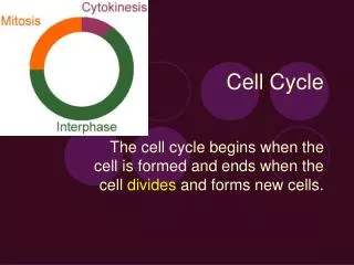
M G2 G1 S Cell cycle - phases • The cell-division cycle is usually divided into four distinct phases. • G1 (gap1) is a growth phase that occurs before • S (synthesis) phase — the stage of DNA replication. This is followed by • a second gap phase, G2, • which precedes M (mitosis) phase, during which chromosome segregation and cell division occurs. R
M R G2 G1 S Cell cycle - driven by cdk´s • Orderly progression through these cell-cycle phases is controlled by the sequential activation of the Cdks. • Cyclines and cyclin-dependent kinases (cdk) • cyclines+ cdk cell cycle-dependent variations in the activity of the kinases phosphorylation of nuclear factors such as RB changes during the cycle • The subsequent phases are controlled by cyclin-cdk pairs as shown below • Cellular stress activation of checkpoint pathways cell-cycle progression is disrupted • The R-point: “restriction point” 2/3 into G1
Cyclin E Cyclin A Cyclin B Cyclin D G0 G1 S M G2 Cyclins • Cyclines and cyclin-dependent kinases (cdks) • The cyclines have oscillating levels during cell cycle • The cyclines are regulatory subunits of the CDK-kinases • cyclines+ cdk cell cycle-dependent variations in the activity of the kinases determined by mitogenic growth factors
Restriction point of the cell cycle • Growth factors (both positive and negative) exert their effect during the G1 phase. • Beyond the restriction (R) point = committed • The restriction (R) point defines a critical time in late G1 after which a cell is committed to undergo DNA replication and is no longer sensitive to growth-factor signalling. After the R point, cell cycle progression can only be halted by conditions of cellular stress, such as DNA damage or mitotic-spindle defects. • Before the restriction point, the cell has a choice between cell division (growth) by continuing the cell cycle, and rest by going into G0 • Beyond the restriction point the cell is commited to proceed until cell division (M) Growth factor sensitive Committed - insensitive
Regulatingcell cycle • Cdk regulation • cyclins, • inhibitory and activating phosphorylation events, • association/ dissociation of inhibitory molecules called Cdk inhibitors (CDIs). • Mitogenic growth factors • exert their effect by promoting the synthesis of the D-type cyclins. • cyclin E is triggered by internal signalling • the appearance of Cdk2–cyclin E kinase activity seems to be synonymous with the restriction point. • The ordered activation of the remaining Cdk–cyclin complexes seems to be self-regulating: • each Cdk–cyclin complex triggers the activation of the next Cdk–cyclin species.
M G2 G1 S RB is active only within a limited time window during the cell cycle • Before the R-point in G1: Rb = hypophosphorylatedactive repressor of growth (inhibits cell cycle progression) • SDS-PAGE: 110 kDa • After the R-point in G1: Rb = hyperphosphorylatedinactive repressor of growth (facilitates cell cycle progression) • SDS-PAGE: 112 - 116 kDa • Rb is dephosphorylated at the end of mitosis • Coupling phosphorylation status/function • Oncoproteins from DNA tumour virus bind/inactivate pref hypo-RB • Only hypo-Rb bind/inactivates andre cellulære proteins/TFs • Stimuli that enhance Rb phosphorylation facilitate proliferation active repressor Rb R Rb P P P P P P Inactive repressor
Gate-keeper model for RB • The R-point functions as a door that is kept closed by Rb • G1 arrest upon overexpression of Rb • Under conditions favourable for proliferation Rb phosphorylated R-door is opened • In cells with lost Rb-function the door is left open all the time • Such cells will also have lost the ability to respond to growth-promoting/-inhibitory signals • Mitogenes (+), TGF (-), contact-inhibition (-) • Two key elements in this model: • upstream signals Rb´s phosphorylation status • Rb´s phosphorylationsstatus downstream effects • Rb as “signal transducer” • Cell cycle-clock RB´s phosphorylation status • RB´s phosphorylation status transcription apparatus involved in proliferation
Cdk4/6 Cyclin D Rb R M G2 G1 S E2F released S-phase genes expressed Gate keeper model
M G2 G1 S Cell cycle clock RB´s phosphorylation status • Multiple Ser/Thr sites in RB are phosphorylated • multiple kinases converge on RB • Multiple sites typical CDK sites • Cyclin D most involved in RB phosphorylation • G1-Cyclins D1, D2 and D3 are regulators of CDK4 and CDK6 • The D cyclins form physical complexes with RB • Regulators which inhibit CDK4/6 will block RB phosphorylation • Cyclin E-CDK2 also contributes to RB phosphorylation • Ectopic expression of cyclin E RB phosphorylation • cyclin E increases significantly towards the end of G1 • viral oncoproteins which block cyclin D binding do not abolish RB phosphorylation Cdk4/6 +cyclin D Rb R Cdk2 +cyclin E
Cell cycle-watch RB´s phosphorylation status • Expression of RB in yeast normal RB phosphorylation requires two types of cyclins • requires two different G1 cyklines: CLN3 + (CLN1 or CLN2) • ∆ CLN3 RB´s phosphorylation normalized by introduction of mammalian cyclin D1 • ∆ CLN1/2 RB´s phosphorylation normalized by introduction of mammalian cyclin E • Different models for cooperation of D and E cyclins • cyclin D-CDK4/6 formation of hyperphosphorylated RB, while cyclin E-CDK2 maintenance of hyperphosphorylated RB • cyclin D-CDK4/6 formation of partially phosphorylated RB better substrate for cyclin E-CDK2 formation of hyperphosphorylated RB • Continuous turnover of phosphate • t1/2 for phosphate on RB ≈ 15 min (due to phosphatase activity) maintenance of phosphorylated status necessary
RB as an integrator of positive growth signals • general: physiological signals that promote proliferation enhanced RB phosphorylation • Growth factors/mitogenic signals receptor intracellular signalling pathways RB phosphorylation cell cycle progression/proliferation • Abundance of extracellular mitogenes sensed as [cyclin D1] • sufficient D1 RB phosphorylation • low D1 RB unphosphorylated
E2F liberated by Rb inactivation • Rb excert its effects through E2F TFs Rb = inactivated Rb = active repressor R-point E2F = activated!
RB´s phosphorylation status = a signal to the trx apparatus • Hypophosphorylated RB binds and inactivates the transcription factor E2F/DP • Hyperphosphorylation of RB E2F/DP liberated and free to activate genes necessary for proliferation
Repressor-mechanism: through chromatin • mechanism for repression • E2F binds DNA ± RB • RB acts as an active repressor associated with DNA-bound E2F • RB recruits HDAC-complexes that cause repression
Repression in several stages • 1. Blocking TAD • 2. Recruitment of HDAC • 3. Recruitment of HMT
Local repression by RB:first deacetylation, then methylation Step 1: deacetylation Step 2: methylation
RB´s Pocket-domain important The Nine Residues Of Papilloma Virus E7 Peptide Contain The LxCxE Motif • Pocket-properties • HDAC1 binds to Rb´s pocket-domain (379-792) • The repressor-function of Rb is located to the pocket-domain • Pocket also bindingsite for viral oncoproteins via LxCxE-motif • All disease related mutations located to the pocket-domain • Model • Rb-HDAC1 association interrupted and Rb´s repressor-function lost when • 1. Rb is phosphorylated • 2. Pocket domain mutated • 3. Virale oncoproteins bind pocket
Rb related pocket proteins • 3 members in the “pocket”-family: RB, p107, p130 • Common: A + B domains forming the “pocket” domain • all natural Rb mutations in A or B • similarities in cell cycle-dependent phosphorylation • Unequal with regard to associated cyclins and expression • Few or no mutations in p107 and p130 found in human cancers • Parallel controls through several “pocket-proteins” and multiple E2Fs • RB binds E2F-1, 2 and 3 • p107 binds E2Fs 4 • p130 binds E2Fs 4 and 5 • different E2Fs have different functions (se below)
E2F liberated by Rb inactivation • Rb excert its effects through E2F TFs Rb = inactivated Rb = active repressor R-point E2F = activated!
The E2F/DP-family of transcription factors • E2F/DPs = a group of bHLH-ZIP factors • E2F/DP - heterodimers of E2F + DP • E2F: 6 distinct related TFs (E2F-1-6) • DP-partners: 2 TFs (DP-1, DP-2) • All possible combinations • 3 subgroups • Activating E2Fs • Potent activators • Repressive E2Fs • Active repressors • E2F6 - repressor? • Pocket independent • Ass polycomb-complex
Target genes controlled by activating E2Fs • E2F sites • common konsensus binding site: TTTCCCGC • optimal binding to TTTCGCCGCCAAAA (to motsatt orienterte overlappende sites) • No difference in sequence preference between different E2Fs • target genes: E2F controls the transcription of cellular genes that are essential for cell division: • cell cycle regulators • such as cyclin E, cyclin A, Cdc2, Cdc25A, RB and E2F1, • enzymes that are involved in nucleotide biosynthesis • such as dihydrofolate reductase, thymidylate synthetase and thymidine kinase • the main components of the DNA-replication machinery • Cdc6, ORC1 and the minichromosome maintenance (MCM) proteins. • E2F knock-out - a paradox
The activating E2F1, E2F2 & E2F3 • Key role: the activation of genes that are essential for cellular proliferation and the induction of apoptosis. • Overexpression →proliferation • quiescent cells →re-enter the cell cycle • Override various growth-arrest signals • Transformation of primary cells • Knock-outs →reduced proliferation • E2f3-/- MEFs: defective in the mitogen-induced activation of almost all known E2F-responsive genes • the combined mutation of E2f1, E2f2 and E2f3 is sufficient to completely block cellular proliferation.
The activating E2F1, E2F2 & E2F3apoptosis ?? • Key role: the activation of genes that are essential for cellular proliferation and the induction of apoptosis. • The threshold model of the activating E2Fs. • The activating E2Fs contribute to a pool of E2F activity. Once this reaches a critical level, it triggers proliferation (threshold 1) or apoptosis (threshold 2).
The ‘activating’ E2Fs are key targets of RB • E2F1-3 interact specifically with RB • The ‘activating’ E2Fs are specifically regulated by their association with RB, but not with the related pocket proteins p107 or p130. • RB binds transactivation domain (TAD) in E2F • Release from Rb is triggered by the phosphorylation of RB in late G1 and correlates closely with the activation of E2F-responsive genes. • The functional inactivation of RB induces the same phenotype as the overexpression of E2F: • inappropriate proliferation, p53-dependent and p53-independent apoptosis • Mutation of either E2f1 or E2f3 in RB-deficient embryos is sufficient to suppress all these defects. Rb binding
The repressive E2F4 & E2F5regulated in a different fashion • Significant levels of E2F4 and E2F5 are detected in quiescent (G0) cells, • E2F1, E2F2 and E2F3a are primarily restricted to actively dividing cells. • The E2F subgroups bind to different pocket proteins. • Whereas the activating E2Fs are specifically regulated by RB, E2F5 is mainly regulated by p130, and E2F4 associates with each of the pocket proteins at different points in the cell cycle. • E2F4 is expressed at higher levels than the others, • it accounts for at least half of the RB-, p107- and p130-associated E2F activity. • The subcellular localization of the endogenous E2F4 and E2F5 complexes is also regulated, • E2F1, E2F2 and E2F3 are constitutively nuclear, whereas E2F4 and E2F5 are predominantly cytoplasmic. In complex with pocket proteins nuclear. • KO: ‘repressive’ E2Fs are important in the induction of cell-cycle exit and terminal differentiation.
Cell-cycle regulation of individual E2F complexes • The spectrum and subcellular localization of the E2F–complexes from G0 to the restriction point (late G1). The approximate abundance of each complex is indicated by their relative size. Active repression of target genes Repressive Complexes Replaced With Acitvating ones Derepression + activation of target genes Cell cycle
E2F/DP only active in a window of the cell cycle (late G1 early S) • Early G1: active RB E2F/DP turned OFF • The R-point: inactivated RB E2F/DP turned ON • E2F/DP liberated → activation of E2F-dependent promoters • Late S: E2F/DP turned OFF again • cyclin A/cdk2 → phosphorylation of E2F/DP → reduced DNA-binding → target genes turned off
EF26 - another mode of repression • Less well studied
Other effector-functions of RB • RB is abundant in the cell • RB/E2F ≈ 100 • RB can bind opp a range of proteins other than E2F • consensus binding motif: LxCxE • TFs: Elf-1, MyoD, PU.1, ATF-2 • nuclear tyrosine kinase: c-Abl • hypo-RB binds catalytic domain inactivates kinase • By binding up several different effector-proteins → coordinated control of several downstream growth-related pathways • Still - the E2F-pathway plays a key role • Ectopic expression of E2F → overrides RB-block
RB as integrator of negative growth inhibitory signals • general: physiological signals that inhibit proliferation → reduced RB phosphorylation → cell cycle don’t pass R • acts indirectly through CDK-inhibitors (CDKIs) → reduced CDK activity → reduced RB phosphorylation • Three well known physiological growth inhibitory signals • TGF • cAMP • contact inhibition • TGFb growth inhibtion: 3 mechanisms • TGF→ posttranslational modification/activation of CDKI p27Kip1→ inactivation of CDK2,4 and 6 → reduced RB phosphorylation • TGF→ induction of CDKI p15INK4B→ inactivation of CDK4 and 6 through cyclin D competition → reduced RB phosphorylation • TGF→ reduced level of CDK4 → reduced RB phosphorylation
RB as integrator of negative growth inhibitory signals • cAMP/contact inhibition / growth inhibition • cAMP → mobilize CDKI p27Kip1→ inactivation of CDK2,4 and 6 → reduced RB phosphorylation • Irradiation/DNA-damage • DNA-damage → enhanced p53 → induction of CDKI p21Waf1/Cip1→ inactivation of CDK4 and 6 → reduced RB phosphorylation → G1 arrest → time to repair DNA
RB and cancer - several ways of killing RB-mediated cell cycle control • Rb mutation • retinoblastoma, small cell lung carcinomer, sarcoma, kidney carcinomas • RB inactivated by RB-binding oncoproteins • cervical carcinomas: human papillomasvirus E7 oncoprotein • amplification of cyclin D genes • esophageal-, bryst- and squamous cell carcinomas • in B-cell lymphomas due to chromosome translocation • Virus-encoded D-type cyclins • Herpesvirus saimiri • amplification of the CDK4 gene • glioblastomas • gliomas • deletion of genes for p15 or p16 • several carcinomas • also germ-line mutations in familial melanomes I alle cases: lost RB function open R-door free E2F cell cycle without brakes