Download
1 / 26
260 likes | 335 Views
Delve into the fascinating world of microorganisms, from beneficial bacteria to harmful pathogens. Learn about microbiology's various specializations and the classification of different types of cells. Explore the structures and functions of bacteria, including cell walls and genetic composition.

E N D
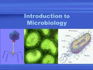
Introduction To General Microbiology Prof. Dr. Asem Shehabi Faculty of Medicine University of Jordan
The Microbial World • The microbial world is composed of commensally and pathogenic Microbes/ Microorganisms..Bacteria, Fungi (Yeast/ Moulds), Algae, Protozoa/ Parasites and viruses. • Microbiology is concerned with the study of these microbes..Mostly are beneficial.. Few species cause harmful effects ..disease in human & animals. • Microorganisms are unicellular cell.. too small to be seen with the naked eye.. recognized by light microscope .. Bacteria, fungi & Parasites ..their sizes above > 0.1 um • Most microbes capable of grow & existence as single organism or together with others .. Widely distributed in Human, Animal, Nature.
Microbiology • Viruses sizes < 0.01um Composed of only DNA or RNA..grow only in living cells/tissue culture.. are non independent cellular entities..can’t be considered true microorganisms..Their presence structures can be seen only with electron microscope. • Microbiologyhas many areas of specialization including Bacteriology, Mycology (fungi), Virology,Medical microbiology, Immunology, Food microbiology, Biotechnology, Microbial genetics ..Industry.. Agriculture Veterinary.
Classification of Microorganisms • Two fundamentally different types of cells are classified in the microbial world,Prokaryotic .. Eukaryotic cells. • Eukaryotic cells have a"true" nucleus.. Prokaryotic cells have a naked nucleus.. composed of a single DNAChromosome.. not enclosed within a nuclear membrane. • The shape of Prokaryotic cells.. Bacterial, Fungi ,Parasites cells are of fundamental importance in the classification and identification of these microbes in Labs.
Bacteria • Bacteria are unicellular microorganisms..Size (0.2umDiameter, 0.2-10um Length)having a variety of shapes ..Growth patterns & metabolic characteristics allowing their classification. • Major bacteria cell shapes are arranged:Coccus/cocci, Bacillus/bacilli or Rods, Coccobacilli, Spiral forms- spirochetes, Vibrios • Individual cells may be arranged in pairs or clusters or chains.. Their morphologies are useful for the identification & classification of bacterial Genera and Species.. colored by Gram-stain or other stains (Fig-1)
Bacterial Cell structures-1 • Cell wall structures:A rigid cell wall, composed of many peptidoglycan layers .. outer membrane, A periplasmic space, a cytoplasmic membrane lacking sterols, Cytoplasma ..70S ribosomes, mesomes, storage granules -Lipids, glycogen, polysaccharides, sulfar, phosphate .. Others storage compounds. • Bacterial genome.. One single supper coiled DNA chromosome, plasmids(>1). • Flagella:Organs of motility, composed of flagellins (polymer proteins) long filament.. length up to 20 um (Figure 2).. Attachment.. Nutrition..Single polar flagellum (monotrichous).. Several polar flagella at one, each end of the cell or covering the entire cell surface (peritrichious).. antigenic determinants (H-antigen)..observed during bacterial infection.
Bacterial Cell structures-2 • Fibmriae.. Pili:Small surface appendages (protein).. Few numbers Pili.. Sex function /Large Numbers fimbriae..specific functions .. Attachment/Adhesion to host epithelial cells/colonization & antigenic determinants. • Capsules:surface layer of cell wall.. a slime layer composed mostly of high molecular weight polysaccharides.. provide resistance to phagocytosis.. avoid the killing effects of lysosomal enzymes, and serve as antigenic determinants.. (K-antigen).. Major virulence factor in certain bacteria • Virulence factor.. Any bacterial part/product Associated with pathogenic potential.. causing human/animal infection/disease.
Bacterial Cell wall Structures-1 • Bacterial cell wall contains a special polymer called Peptidoglycan. Its basic structure is a carbohydrate backbone of alternating units of N-acetyl glucosamine and N-acetyl muramic acid. • These are cross-linked with oligopeptides.. contain both D- and L-amino acids. • Teichoic acid-Lipoteichoic acids:found only in Gram-positive bacteria. • Lipopolysaccharides:Lipopolysaccharides (LPS) found only in Gram-negative bacteria.
Bacterial Cell wall Structures-2 • LPS structures are composed of lipid A,which binds to the outer membrane.. Endotoxic portion of the molecule.. Causing Toxic Shock.. High Fever, Sepsis • The polysaccharide moiety appears on the cell surface, serving as an antigenic determinant O antigen- Host cells develop during bacterial Infection..Anti-O AB • Cell wall is the basis for classification of bacteria into Gram-positive & Gram-negative by Gram-stain • Cell membrane: A phosolipid bilayer responsible for transport of ions, nutrients and waste across the membrane.. Control the cell plasma contents
Gram-Stain A- Gram-positive bacteria have a thick layer of peptidoglycan, Many sheets.. external to the cytoplasmic membrane.. Lipoteichoic acids.. stained Blue.. Staphyloccocus, Streptocooci, Bacillus..Protoplasts..L-form..Lysozyme effect..Loss Most Cell wall, Burst +Lysis B- Gram-negativebacteria contain lipopolysaccharide (LPS) attached to the outer membrane... source of the O-antigen and endotoxin reaction.. Stained Purpel/Red.. Enteric bacteria group.. Esch. coli, Klebsiella, Salmmonella Pseudomonas, * Spheroplasts
Spore-Forming Bacteria • ENDOSPORE FORMATION: The process of sporulation begins when vegetative (actively growing cells) exhaust their source of nutrients .. begin of forming endospores.. Common in nature (Figure 4). • Spore forming Bacteria are very resistant to lysozyme, heat, radiation, drying and can remain dormant for hundreds of years in nature.. Once conditions are again favorable for growth, the spores can germinate and return to the vegetative state. • Aerobic Bacillusgroup & Anaerobic Clostridium..developEndospore formation.. Both are widely distributed in nature ..intestinal -human and animals.
Growth & Nutrition-1 • Requirements for bacterial growth:oxygen, water, pH, temperature, source of carbon, nitrogen ( organic compounds), inorganic salts.. Na, K, S, P, Ca, Mg, Cl, Fe, vitamins, etc. • Obligate Aerobic bacteria..M. tuberculosis, P.aeruginosa grow using respiration.. oxidation.. recipientOxygen.. Aerobic bacteria encounter the oxygen damage during their growth by producing oxidizing enzymes: • Peroxidase:Oxidize H2O2 into 2H2O+NAD. • Superoxidase dismutase:Reduce O2- into H2O2 +O2 .. • Catalase:Reduce H2O2 into 2H2O+O2.
Growth & Nutrition-2 • Certain Pathogens grow with reduced level of oxygen.. Microaerophilic bacteria..Neisseria spp. • facultative anaerobes.. prefer growing in the presence of oxygen, but can continue to grow without it.. Most human pathogens & normal flora.. Staphylococci, streptococci, E.coli • Obligate Anaerobicbacteria grow by absence of oxygen.. use recipient inorganic molecule.. Fermentation.. Mostly found in intestinal tract (95-99%), Mouth &Vagina(90%) • Examples Anaerobes: Gram-ve Bacteriodes fragillis, G+ve Clostridia, Gram+ve Cocci
Growth & Nutrition-3 • Bacteria classified by the source of their energy oxidation-reduction process into two groups: • Heterotrophs:derive energy from breaking down complex organic compounds.. protein, sugar, fats.. human tissues.. All commensals-pathogens • Autotrophs:fix carbon dioxide to make their own food source.. using light energy photoautotrophic, or oxidation of nitrogen, sulfur, other elements chemoautotrophic.. sulfur & nitrogen fixing bacteria.. Environment. • Saprophytic bacteria/ Nonpathogenic.. take energy by fermentation/respiration.. found in nature.. in decaying material.. soil, water..vegetations..circulation of minerals.
4/ • Culture Media:Nutrients (carbohydrates & proteins, blood, minerals) Source.. Water..Broth medium, Solid medium/ Blood agar, Petri dishes/Plate, Growth/Culture (Fig 5) • Neutrophilic bacteria.. Grow best pH (7-7.2) Most human-animal commensales & pathogens • AcidophilicBacteria < 5 pH.. Lactobacilli • Mesophilic Bacteria (20-40C)..Most human commensal & pathogens. • Psychrophilicbacteria(<10C), Thermophiles bacteria (> 60C)..Common in hot spring water • Counting bacteria growth: Plate counts, Turbidity, Dry weight using solid culture agar
Bacterial growth-1 • Bacterial growth is the division of one bacterial cell into 2 identical daughter cells..4,8.16.. binary fission..Generation time ( 15-25 min), most human commensal & pathogens.. Each produce one colony 103 -109 cells ( Fig-4). • Bacterial Strain originated from a single cell. • Baterial Growth Curve:4 phases of visible growth...Lag, Log, Stationary, death/ decline. • Measurement of bacterial growth followed by: • A) Growth/enumeration of cells by direct cell counting in nutrient broth.. microscopic or counting viable cells/ colony forming unit.. Plate counts/ Electronic counting..using solid culture media..nutrient agar
2/Measurement of Growth • B) Indirect counting of growthin fluid medium.. most probable number by measuring turbidity, wet or dry weight.. G/ml.. Important in study research to detect antibiotics & treatment of infection. • Types of culture media: • General culture media: Nutrient agar, blood agar, chocolate agar..growth of most human pathogens.. Gram-ve & Gram-ve bacteria. • Selective & differential media..MacConkey agar Bile salts+ Lactose+neutral red dye ..Gram-ve bacteria, E.coli, other enteric bacteria • Selective media: S-S agar .. For Isolation of Salmonella, Shigella , V.colerae from stool specimens.