Download
1 / 43
430 likes | 486 Views
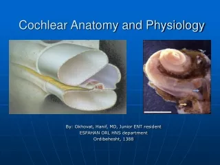
Understanding the intricate structures of the cochlea and how they function in auditory perception. Explore topics such as the osseous labyrinth, membrane components, and cochlear mechanics for a detailed insight into auditory transduction.

E N D
Cochlear Anatomy and Physiology By: Okhovat, Hanif, MD, Junior ENT resident ESFAHAN ORL HNS department Ordibehesht, 1388
Essentials of Anatomy: • Osseous Labyrinth • Modiolus • scala vestibuli : Contains perilymph • Helicotrema • scala tympani: Contains perilymph • scala media(cochlear duct): contains endolymph • perilymph: extracellular-like fluid, high Na+ and low K+ • endolymph: intracellular-like fluid, high K+ and low Na+ Membranous labyrinth: (1) Reissner's membrane (2) the lateral wall: spiral ligament, stria vascularis (3) the basilar membrane: the organ of Corti the organ of Corti contains : supporting cells, pillar cells, outer hair cells, and the inner hair cells • Tectorial Membrane : an acellular, extracellular matrix
Sensory Cells Stereocilia • crucial for hair cell transduction. • Stereocilia: not true cilia long, stiff microvilli • Stereocilia increase in length from the cochlear base toward the apex and laterally across the rows of the outer hair cells. • Tip links occur between the tips of the shorter stereocilia and shafts of the longer stereocilia • Mature cochlear hair cells, unlike vestibular hair cells, do not contain a kinocilium. • The outer hair cell stereocilia bundle forms a V -shaped or W -shaped pattern. • The inner hair cell stereocilia form a slightly curved bundle of three or four rows of stereocilia. • The tips of the stereocilia of the longest row of the outer but not the inner hair cells are attached to the undersurface of the tectorial membrane.
Outer hair cells: 13,400 outer hair cells, cylindrical insert into a cup-shaped Deiters' cell body at their basal pole surrounded by the fluid-filled spaces of Nuel The cells increase in length toward the apex and laterally across the rows Inner Hair Cells differ from the outer hair cells: a single row of flask-shaped cells Instead of W arrangement of stereocilia in OHC, IHC stereocilia have a linear pattern
Chapter 150 – COCHLEAR TRANSDUCTION AND THE MOLECULAR BASIS OF PERIPHERAL AUDITORY PATHOLOGY Cochlear Mechanics: Passive Active
PASSIVE COCHLEAR MECHANICS Fundamental nature of cochlear mechanics is evident even in cadavers Not requiring adenosine triphosphate (ATP) Components of the principal elements underlying passive mechanical transduction: 1)Basilar membrane : the central structure of the end organ both functionally and anatomically. 2)scala vestibuli and scala tympani: filled with perilymph 3)scala media: filled with endolymph 4)tectorial membrane Reissner's membrane : important homeostatic role No role as an element of passive cochlear mechanics.
sound pressure waves are delivered to the scala vestibuli through oval window propagate through the incompressible cochlear fluids at a velocity of 1.5 km/sec the spread of the pressure wave throughout the cochlea is nearly instantaneous. the higher pressure (or lower, depending on the direction of motion of the stapedial footplate) in scala vestibuli relative to scala tympani produces a pressure differential across the cochlear partition that set the partition into motion
How does cochlea have TONOTOPIC properties? 1) the width of the basilar membrane progressively increases toward apex the width at the base is 100 µm and 500 µm near the apex. 2) the number of cells lining the basilar membrane increases along the basoapical length 3) the length of OHCs and stereocilia increase toward apex. 4) the thickness of the basilar membrane and the density of filaments decrease from base to apex the basilar membrane and organ of Corti complex is stiffer and less massive at the base than at the apex The result is an end organ with resonant properties that are continuously distributed from base to apex Basilar membrane filters complex fluid waves into sinusoidal constituents. As a result, high-frequency acoustic events are preferentially transduced in the base, because it is stiff and less massive. The inverse is true in the case of low-frequency acoustic events.
Other physical characteristics of the cochlea : 1)the viscous friction between the basilar membrane and cochlear fluids produces a damping effect that limits its displacement. 2)The traveling wave propagates basoapically, because the stiffness of the cochlear partition decreases longitudinally from the base to the apex as its mass increases. The motion and direction of the wave that travels along the basilar membrane is independent of the location of the vibratory source, because : the stiffer and less massive elements forming the base of the organ of Corti are set into motion before the more compliant and massive sections of the apical cochlea It forms a frequency decomposition system: the space-frequency or tonotopic map
Near the characteristic place for a given stimulus frequency, the pressure wave returns to the basal end of the cochlea through the fluids of the scala tympani, where the flexible round window membrane is displaced in an equal but opposite direction of the stapes footplate
Helicotrema: allowing the fluids in the scala vestibuli and scala tympani to communicate directly acts as an acoustic shunt across the cochlear partition reduces the pressure difference between the scalae produced by very-low-frequency stimulation The size of the helicotrema determines the cutoff frequency of the system large openings shunt pressure waves extending to higher frequencies than do small helicotrema. decrease damage to the cochlea produced by intense low- frequency pressure fluctuations
Brace Yourself IT’S SHOW TIME!
ACTIVE COCHLEAR MECHANICS Why bother with ‘active mechanics’ theory? 1)Traveling wave measured in cochleae from cadavers is so broadly tuned as to be inconsistent with a living individual's ability to discriminate closely spaced frequencies 2)Sharp tuning and high sensitivity are lost in cochleae from cadavers 3)The peak of the traveling wave in living animals is very sharply tuned at low levels of stimulation and exhibits nonlinear growth as sound levels increase 4)Basilar membrane vibration relative to stapes displacement is as much as two to three orders of magnitude greater under low stimulus level conditions than at higher levels
So, cochlear mechanics are nonlinear Active mechanics is a metabolically labile, energy-consuming process the active mechanical event underlying amplification is highly localized. the traveling wave peaks at increasingly apical regions of the cochlear partition with increasing stimulus level, shifting the tonotopic map toward lower frequencies at any given place.
Role of Outer Hair Cells in Active Mechanics Destruction of OHCs causes: 1)diminished cochlear sensitivity 2)Loss of frequency selectivity 3)loss of nonlinear operating properties only 5% of all auditory nerve fibers innervate OHCs though there are at least three times as many OHCs as IHCs These findings suggested that the transmission of sensory information to CNS is not the primary function of OHCs.
when OHCs were lost, with IHCs appearing unaffected, some cochlear functions were affected : 1)cochlear sensitivity reduced 2)frequency selectivity reduced 3)input-output curves acquired a more linear property in the region of OHC damage. 4)two-tone suppression (i.e., the reduction in the response to one tone elicited by the presence of another tone) diminished 5)distortion tones (i.e., the appearance of additional tones not present in the stimulus produced by the interaction of two or more primary tones, such as 2f1-f2 or f2-f1, where f1 and f2 represent the primary tones with f1 < f2) are lost
OAE : evidence that an active, OHC-driven mechanism operates within the cochlea, which is capable of producing energy OCB(olivocochlear bundle) activation altered cochlear output as well as otoacoustic emissions and basilar membrane displacement Outer hair cell motility induced by electrical stimulation of the excised cochlea displaced both the reticular lamina and the basilar membrane.
OHCs contract or elongate at very high rates (up to at least 70 kHz) along the length of their longitudinal axes when depolarized or hyperpolarized, respectively OHCs amplify the displacement of the basilar membraneRecently, the voltage-sensitive motor protein underlying the process was identified and given the name prestinintracellular Cl- ions act as extrinsic charged voltage sensors. Salicylates, which are known to cause temporary hearing loss when administered at high doses, are permeable to the plasma membrane and act as competitive antagonists of Cl-, thereby reducing electromotility and, diminishing sensitivity.
Conversion of Basilar Membrane Displacement to Radial Shearing Forces Reticular lamina: interdigitating heads of the inner and outer pillar cells, the apical surfaces of the phalangeal processes of Deiters' cells, and the apical surfaces of the hair cells through which the stereocilia project Reticular lamina is a relatively rigid structure that moves in concert with basilar membrane motion. The tectorial membrane is rigid structure firmly attached to the spiral limbus and overlies the reticular lamina .
displacement of the basilar membrane results in a radial shearing motion between the reticular lamina and the tectorial membrane, a motion that serves as the mechanical trigger of transduction currents The tallest row of stereocilia protrudes from the apical surface of outer hair cell and embeds within the tectorial membrane the shearing motion between the reticular lamina and the tectorial membrane causes stereocilia to bend in the direction of the modiolus or the spiral limbus, depending on whether the basilar membrane is displaced toward the scala tympani or scala vestibuli, respectively. In contrast to OHCs, the stereocilia of IHCs do not appear to firmly contact the tectorial membrane Therefore, during basilar membrane vibration, the mechanical stimulus to IHC stereocilia is the flow of endolymph within the subtectorial space(the channel between the reticular lamina and tectorial membrane).
Radial Displacement Patterns of the Basilar Membrane The foot of the inner pillar cell lies near the bony spiral lamina The foot of the outer pillar cell lies over the basilar membrane and is not supported by bone. When the basilar membrane is displaced in a passive cochlea, movement occurs maximally near the foot of the outer pillar cell When OHCs contract, the reticular lamina pivots at the apex of the tunnel of Corti, and the basilar membrane and reticular lamina are pulled together, enhancing overall basilar membrane displacement. Greatest motion in active cochlea is in the domain of the OHC
HAIR CELL TRANSDUCTION and stereocilia approximately 3500 IHCs and 12,000 OHCs Stereocilia: not true cilia more appropriately: specialized microvilli kinocilia are on the surface of immature auditory hair cells of mammals but are lost during the final stages of differentiation. Approximately 50 to 70 stereocilia protrude from each IHC and about 150 from each OHC
Toward Apex: 1)the number of stereocilia on OHCs decreases to less than half 2)the lengths of the stereocilia of both IHCs and OHCs increase 3)The lengths of OHCs increase, whereas the lengths of IHCs remain fairly consistent Individual hair bundles are organized into three rows of stereocilia Arranged linearly in the case of IHCs In the form of a "W" in the case of OHCs, with the base of the "W" facing the lateral wall of the cochlea
Stereocilia within each row are approximately the same height The longest row is located on the side of the cell that is proximal to the spiral ligament The length of stereocilia decreases in an orderly manner in rows that are located progressively proximal to the modiolus, producing a stairstep configuration The stereocilium tapers near its point of insertion into the surface of the receptor cell The stereocilia pivots, as opposed to flexing, near the surface of the receptor cell, acting like a stiff lever when mechanically distorted.
Hair Bundle Deflections and Receptor Potentials Magnitude of the receptor potential is proportional to the degree of stereociliary deflection in the most sensitive region of its operating range Saturates with larger deflections in either direction. When the bundle is deflected toward the tallest stereocilia, the hair cell depolarizes, Deflections of the bundle in the direction opposite the tallest stereocilia hyperpolarize the cell Displacement-voltage curve is highly asymmetric relative to its resting state responses saturate with smaller deflections directed away from the taller stereocilia than in the excitatory or depolarizing direction Deflection of the hair bundle in a direction perpendicular to this axis produces no response.
Hair Cell Transduction Channels The latency of hair cell response to displacement: 40 μsec Activation time is inversely related to the magnitude of bundle displacement enzymatic systems or second messengers are too slow to underlie the transducer response transduction channels operate in either an open or a closed state, with the probability of being in one or the other state depending on the degree of hair bundle deflection. the asymmetric displacement-voltage curve described previously is explained by the fact that a small population of transduction channels, roughly 10% to 15%, is in the open state at rest. transduction channels are situated near the tips of the stereocilia Each stereocilia has one transduction channels and transduction channels are present in stereocilia of all heights within a bundle
Gating of Transduction Channels tip links stretch between the distal tip of each stereocilium with the side of the longer adjacent stereocilium transduction channels are located at each end of the tip link When the bundle is deflected toward the taller stereocilia, tip links are stretched and physically open transduction channels, causing excitation When the bundle is deflected away from the taller stereocilia, tip links relax, causing the channel to close. Movement of the bundle perpendicular to the axis of symmetry produces little or no response
Acoustic overstimulation can cause tip link breakage Tip links broken as a result of exposure to BAPTA regenerate within 24 hours, restoring mechanoelectrical transduction in the process. Perhaps a similar dynamic occurs in the case of recovery from temporary threshold shifts associated with acoustic overstimulation.
Transduction Channel Ion Selectivity Hair cell transduction channels are nonselectively permeable K+ carries most of the receptor current When transduction channels open, K+ in the endolymph is flushed down a large electrochemical gradient This inward transducer current flows across the basolateral membrane, producing the receptor potential and voltage-gated Ca2+ channels are activated, resulting in an influx of Ca2+ and release of neurotransmitter from the base of the hair cell.
Chapter 149 – COCHLEAR ANATOMY AND CENTRAL AUDITORY PATHWAYS Osseous Labyrinth: surrounds the internal membranous labyrinth Modiolus: internal bony tube communicates with the endolymphatic compartments by way of the ductus reuniens.
Membranous Labyrinthcochlear duct is divided into three regions: (1) Reissner's membrane (2) the lateral wall ,which includes the spiral ligament, stria vascularis, spiral prominence, and the external sulcus (3) the basilar membrane and osseous spiral lamina, includes Claudius' cells; Boettcher's cells; the organ of Corti;inner sulcus; and spiral limbus. the organ of Corti contains : Hensen's cells, Deiters' cells, pillar cells, inner border cells, outer hair cells, and the inner hair cellsspiral limbus contains : the interdental cells and overlying tectorial membraneRosenthal's canal contains : the spiral ganglion and communicates with the modiolus
(1)Reissner's Membrane (vestibular membrane) a three-layered structure. Attached medially to the modiolar edge of the spiral limbus and laterally to the spiral ligament at the apical edge of the stria vascularis. The cells facing endolymph have a low-cuboidal shape, exhibit numerous apical microvilli, and are sealed along their lateral borders by tight junctions. The cells facing perilymph have a squamous shape and are loosely joined together. Reissner's membrane seems to be freely permeable to water, but it restricts paracellular passage of molecules into the endolymphatic space by tight junctions. hydrops : In certain pathologic conditions (i.e., Ménière's disease), Reissner's membrane bulges out into the space of the scala vestibuli.
(2)Lateral Wall A. Spiral Ligament greatest portion of the lateral wall of the cochlear duct consists of cells rich in ion-transporting enzymes. lateral boundary: inner surface of the otic capsule medial boundary: formed by the stria vascularis and the spiral prominence. extends into the scalae vestibuli and tympani and forms a lateral route of communication between these two perilymphatic channels. The cells of the spiral ligament : recycling of K+ from endolymph through the hair cells and into to the stria vascularis(source of endolymphatic K+) Anchoring cells: exert tension on the basilar membrane,at the junction of the spiral ligament and the otic capsule.
B. Stria Vascularis forms the endolymphatic border of the cochlear duct. extends from the attachment of Reissner's membrane to the spiral prominence. lacks a basement membrane. three cell types (marginal, intermediate, and basal cells) and intraepithelial capillaries. The marginal cells : primary functional units of the stria vascularis and produce the positive DC endocochlear potential and the low-sodium, high-potassium ion concentration of cochlear endolymph. The intermediate cells: melanocytes, phagocytic activity, contain carbonic anhydrase enzyme. C. Spiral Prominence D. External(outer) Sulcus : contain carbonic anhydrase.
(3)Basilar Membrane from the lateral edge of the osseous spiral lamina basilar crest : wedge shaped tissue, insertion of basilar membrane into the spiral ligament spiral length averages 31.5 mm. width increases toward the cochlear apex from 150 to 450 µm. width divided into : 1)pars arcuata(medial) 2)pars pectinata(lateral) the thickness of the pars pectinata decreases toward the apex Changes in the width and thickness of the basilar membrane along its length are responsible for the propagation of the traveling wave and the frequency-specific maximal vibrations of the membrane. vas spirale: A spirally directed vessel ,beneath the inner tunnel of Corti, rudimentary exchange of metabolites of the organ of Corti : by means of diffusion of perilymph through the basilar membrane and spaces of Nuel.
A. Claudius' Cells Claudius' cells form a tight cellular border between endolymph of the scala media and perilymph of the scala tympani. B. Boettcher's Cells Boettcher's cells occur most frequently in the cochlear base decrease in number completely toward the apex. to produce fibronectin and other matrix components for the basilar membrane. contain carbonic anhydrase. C. Organ of Corti primary function: convert mechanical vibrations to neural impulses recent evidence : hair cells are able to mechanoelectrically tune the output of the organ of Corti. lateral to medial components: Hensen's cells, outer tunnel of Corti, three to four rows of outer hair cells, Deiters' cells with phalangeal processes, spaces of Nuel, outer pillar cells, inner tunnel of Corti, inner pillar cells, inner hair cells, inner phalangeal cells, inner border cells . reticular lamina : A rigid platelike structure formed by the apical processes of the supporting and sensory cells of the organ of Corti. Toward the cochlear apex, increases occur in : 1)the radial area of the organ of Corti, 2)the length of the inner and outer hair cell bodies and their stereocilia, 3)the angle and width of the apical surface of the organ of Corti relative to the basilar membrane, 4)the length of the pillar cell head plates, 5)the height of Hensen's cells.
Supporting Cells Hensen's Cells not part of the reticular lamina. tectal cells : Hensen’s cells that do not reach the endolymphatic space. outer tunnel of Corti : a fluid filled space Between the last row of outer hair cells and Hensen's cells. Deiters' Cells Deiters' cells support the outer hair cells at their base and apex. phalangeal process: a thin process containing microtubules and filaments,, branches off the Deiters' cell body and extends obliquely to the apical process of adjacent outer hair cells. reticular lamina : This dumbbell-shaped apical pole joins together four different outer hair cells in a rigid platelike array called the reticular lamina. spaces of Nuel: The fluid-filled spaces between the outer hair cell bodies and the phalangeal processes of the Deiters' cells.
Pillar Cells tunnel of Corti : The inner and outer pillar cells oppose each other and form tunnel of corti. the reticular lamina. Each pillar cell has a broad base. The basilar membrane, spaces of Nuel, and the inner tunnel of Corti probably contain perilymph Inner Border and Phalangeal Cells The inner border and phalangeal cells separate the inner sulcus cells from the medial surface of the inner hair cells. The inner border cells form the most medial edge of the organ of Corti. Basally, the inner phalangeal cells envelop unmyelinated nerve fibers associated with the inner hair cell.
Inner Sulcus spirally directed open channel bounded by the lateral edge of the spiral limbus, by the medial edge of the organ of Corti, and apically by the tectorial membrane. Spiral Limbus spirally directed shelf of vascularized connective tissue that overlies the medial region of the osseous spiral lamina. Reissner's membrane is attached at its most medial edge. Its lateral edge forms a wedge-shaped prominence called the tooth of Huschke The endolymphatic surface of the spiral limbus is covered by a thin extracellular matrix called the limbal portion of the tectorial membrane. Directly beneath the limbal portion of the tectorial membrane lie interdental or T cells. They are T -shaped cells whose apical surface forms broad plates across the apical surface of the spiral limbus.
Tectorial Membrane an acellular, extracellular matrix overlies the spiral limbus, inner sulcus, and the organ of Corti. fibrous materials, hydrated by endolymph. divided into six regions: limbal layer, fibrous matrix, marginal band, cover net, Hensen's stripe, and Hardesty's or Kimura's membrane. main protein of the tectorial membrane: Type II collagen the cover net : On the endolymphatic surface, radial strands of material extend out toward the margin of the membrane in an oblique apical direction. the marginal band : The lateral margin of the membrane forms a discontinuous net of material Hardesty's membrane : That portion of the tectorial membrane that faces the organ of Corti, a thin, poorly defined region of material which overlies the outer hair cells Osseous Spiral Lamina spirally directed shelf of bone extends from the modiolus to the medial attachment of the basilar membrane The interior of the spiral lamina forms a channel and a pathway for the nerve fibers that travel to and from the organ of Corti. broad in the cochlear base and narrows toward the apex. At its lateral margin, the bone becomes thinner and perforated by channels called the habenulae perforata, where nerve fibers lose myelination and enter the organ of Corti
VASCULAR SUPPLY The labyrinthine artery : enters the internal auditory meatus with cranial nerve (CN) VIII as a branch of the anterior inferior cerebellar artery. The labyrinthine artery branches to form common cochlear artery and anterior vestibular artery. the common cochlear artery gives off two branches: the spiral modiolar artery: travels apically the vestibulocochlear artery: vascularizes the lower basal cochlear turn. The spiral modiolar artery gives off two coiled branches (i.e., the radiating arterioles) that travel apically over the scala vestibuli and basally to vascularize the spiral ganglion and spiral limbus. The cochlea is drained by the spiral modiolar vein