References
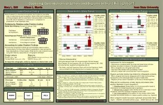
BRCA1 mutation detected by CEL I truncation assay using native 2% agarose gel. Multiplexing five 490 bp PCR products and reacting with CEL I. BRCA1 Exon 11 Section 4 (2014 to 2500 nt in BRCA1 gene). Polymorphism detection in the MED1 gene of 100 patients

References
E N D
Presentation Transcript
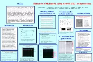
BRCA1 mutation detected by CEL I truncation assayusing native 2% agarose gel Multiplexing five 490 bp PCR products and reacting with CEL I. BRCA1 Exon 11 Section 4 (2014 to 2500 nt in BRCA1 gene) Polymorphism detection in the MED1 gene of 100 patients Three samples are pooled for each reaction GIRAFF Genomic Isogenicity Review by Annealing of Fractionated Fragments bases Detection of Mutations using a Novel CEL I Endonuclease 1 2 3 4 5 6 7 8 9 10 Abstract NEGATIVE CONTROL: 5 products multiplexed, none containing any polymorphisms , none containing any mutations • LaneSample • 100 base marker • 1kb marker • exon 2 Control • exon 2 AG deletion • exon 20 Control • exon 20 C insertion • exon 11.9 Control • exon 11.9 4bp deletion • exon 11.4 Control • exon 11.4 G-A change • DNA was visualized by staining with Sybr Green. M P 427 bp PCR product POSITIVE CONTROL for Mutation: Mutation at 2154 nt, (green peak at 347 bases), plus one polymorphism at 2430 nt. (blue peak at 414 bases) Anthony T. Yeung 1, C. A. Oleykowski 1, S. Griffith 1, D. Besack 1, A. Godwin 1, E.V. Sokurenko 2, S. Henikoff 3, and E. Nicolas 1. 1 Fox Chase Cancer Center, 2 U. of Washington, Seattle, 3 Fred Hutchison Cancer Center CEL I cut at an A9 stretch in the intron P P P P P M Lanes 1-34 are 100 BRCA1 patient DNA samples in pools of 3, treated with CEL I. Lanes 34 and 35 have no CEL I to show PCR background. Pools 3, 9, 14,17, 18, 20, 22-26, 29, 30, and 32-34 contain at least one polymorphism allele. Many genes such as p53, BRCA1, BRCA2, PTEN, and APC, that are often mutated in cancer exhibit a large variety of mutations. The CEL I mismatch endonuclease discovered in our laboratory (Nucleic Acids Res. 26, 4597-4602, 1998) is a powerful tool for screening for unknown mutations in the DNA of patients. The enzyme is compatible with a variety of manual or automated methods of fragment analysis, from native agarose gels to automated DNA sequencing-based GeneScan, in denaturing gels or capillary electrophoresis. The assay requires no optimization for PCR fragments of less than 500 bp. Other applications of this enzyme include screening of plant and zebra fish genes for chemically induced mutants as a replacement for gene knockouts, and in the genomic scanning of antibiotic resistance mutations in bacteria. POSITIVE CONTROL for Mutation: Mutation at 2035nt, (green peak at 466 bases), plus 3 polymorphisms (2196, 2201,2430 nt) (blue peaks at 184,187 and 414 bases) (green peaks at 305 and 300 bp) CEL I blue cut CEL I green cut at polymorphism in exon P P P P P Multiplex 5 unknown samples: Results 3 polymorphisms (2196, 2201,2430 nt) (bue peaks at 184,187 and 414 bases) (green peaks at 305 and 300 bp) Detecting multiple mutations in BRCA1 Genomic scan for bacterial mutations P P P PCR primers Multiplex 5 unknown samples: Results 2 polymorphisms ( 2201, 2430 nt) (blue peaks at 187 and 414 bases) ( green peak at 300 bases) Agarose gel assay Basic Scheme CEL I is a glycoprotein from celery, and many green plants. It is highly stable, but can be inactivated with chelation or SDS. It cuts a heteroduplex that contains a base-substitution or a DNA loop at the 3' most phosphodiester bond of the mismatched nucleotides. Initially, the cut is a single-stranded nick. With more enzyme or longer incubation, the single-stranded nick can be converted to a double-stranded truncation, thereby allowing two assay formats. When a DNA heteroduplex is terminal labeled with fluorescence reporters, the single-stranded cut produced by CEL I at mismatches allow the shorter fragments to be detected with denaturing gels at single nucleotide resolution. This method is used in our center to screen for mutations in cancer genes. It is also in use in other laboratories where DNA pools may contain one mutation allele mixed with up to 17 normal alleles. The Tilling procedure (ref. 4) applies CEL I procedure to find EMS induced mutants of specific genes from a library of mutants. On the other hand, the double-stranded cut method allows the use of native gels to visualize the truncated fragments. This method has enabled a Giraff procedure to detect a single mutation in a bacterial genome (ref. 5). Normal allele 5’ ----------------GGCTCACGT-------------------- ----------------CCGAGTGCA-------------------- 5' Conclusions 5’ ----------------GGCTCGCGT-------------------- ----------------CCGAGCGCA-------------------- 5' Mutant allele Mismatch specificity of CEL I is the reverse of that of the post-replication mismatch repair system Data from ref. 5. Panel A shows fragments isolated for forming heteroduplexes. Panel B shows detection of mutation in 7 Kbp fragments by CEL I, using Southern detection with a cosmid of that region as a probe. The probe can be any subset of the genome. PCR with 5’ labeled blue primer and green primer • CEL I mutation detection assays are easy to use for all mutations with good dynamic range for incubation duration and enzyme quantity. • Homozygous mutants are detected by pooling the PCR products of multiple individuals in an assay. • The strength of the assay is in the detection of unknown mutations with positional information. • Detection at single nucleotide resolution increases the signal to noise ratio. • This mutation detection procedure is suitable for full automation when capillary sequencers are used. 5’ ----------------GGCTCACGT-------------------- ----------------CCGAGCGCA-------------------- 5' 5’ ----------------GGCTCGCGT-------------------- ----------------CCGAGTGCA-------------------- 5' HT Screen for plant gene mutants Two forms of heteroduplexes are formed in PCR as well as the original homoduplexes Introduction CEL I cuts at the 3’ side of a mismatch in one strand of a heteroduplex DNA molecule. The truncated blue and green fragments are measured by Genescan fragment analysis. The sum of the two fragments approximately equals the full length of the PCR product. Detection of a mutation in the ARCS gene of steroid sulfatase C Detection of a rare mutation in exon 1(0.005% allele) Research funded by: Department of Defense, U.S. Army Medical Research and Materiel CommandDMAD17-97-1-7286 Full length • CEL I properties suggest that it is not for DNA repair, but for • plant senescence and remodeling • A very stable mannosyl glycoprotein of about 43 KDa, 29KDa is polypeptide. • The strongest activity is at mismatch heteroduplex, but also shows RNase, single-stranded DNase, and exonuclease activity. • DNA incision is at the 3’ side of a mismatch nucleotide, in one DNA strand only per DNA duplex. Incision makes 3’ OH and 5’ PO4. • Neutral pH optimum, stimulated by Mg++ especially for mismatch cutting. Zn++ is required for activity as indicated by inhibition by 1 mM phenanthroline in the presence of 10 mM Mg++. Only Ca++ can substitute for Mg++. References Mutation cut • Oleykowski, C. A., Bronson Mullins, C. R., Godwin, A. K., and Yeung, A. T. Mutation detection using a novel plant endonuclease. Nucleic Acids Research26:4597-4602, 1998. • Kulinski, J., Besack, D., Oleykowski, C. A., Godwin, A. K., and Yeung, A.T. The CEL I Enzymatic Mutation Detection Assay. Biotechniques, 29,44-48, 2000. • Yang, B., Wen, X., Oleykowski, C. A., Kodali, N. A., Miller, C. G., Kulinski, J., Besack, D., Yeung, J.A., Kowalski, D., and Yeung, A. T. Purification, cloning and characterization of the CEL I nuclease. Biochemistry,39, 3533-3541, 2000. • Colbert, T. G., Till, B., Tompa, R., Reynolds, S. H., Steine, M., Yeung, A.T., McCallum, C. M., Comai, L., and Henikoff, S. (2001) High-throughput screening for induced point mutations. Plant Physiology. 126. 480-484 • Sokurenko E.V., V. Tchesnokova, A. Yeung, C. Oleykowski, E. Trinchina, K. Huges, R. Rashid, S. Moseley, M. Brint, and S. Lory. Detection of mutations and polymorphisms in large genomic regions. Nucleic Acids Res. 29: e111, 2001. • Email: Anthony Yeung at AT_Yeung@FCCC.edu • http://web-apps.fccc.edu/fccc/yeung/index.html Data on Tilling from reference 4, Colbert et al.: LICOR 96 lane sequencing gel image of 480 Arabidopsis plants in pools of 5 being screened for EMS chemically induced mutations in a domain of a specific gene. Left panel is the bottom strand and the right panel the top strand of a IR-fluorescence labeled PCR product, respectively. CEL I cut at a heteroduplex in a lane in the bottom strand has a corresponding signal in a lane for the top strand. One technician in one afternoon can discover 5 mutants in a given gene. This method is being used in several plant and zebra fish projects. ABI 373 Genescan image of CEL I mutation detection. 100 patient’s DNA were used to form pools of 3 each and used as template for PCR. CEL I treating the DNA reveals a band in the pool that contained a heteroduplex. Red = size standards.