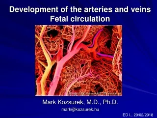
FETAL CIRCULATION
FETAL CIRCULATION. Dr. Tariq Hussain Assistant Professor Pharmacology and Toxicology College of Veterinary and Animal Sciences Jhang. Objectives:. Describe the normal fetal circulation and mention the changes that occur in it at and after birth. Anatomy and Physiology Fetal Circulation.

FETAL CIRCULATION
E N D
Presentation Transcript
FETAL CIRCULATION Dr. Tariq Hussain Assistant Professor Pharmacology and Toxicology College of Veterinary and Animal Sciences Jhang.
Objectives: • Describe the normal fetal circulation and mention the changes that occur in it at and after birth.
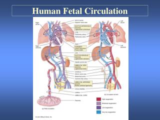
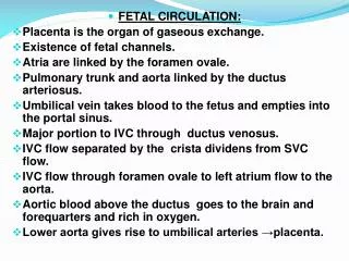
Anatomy and PhysiologyFetal Circulation • Umbilical cord • 2 umbilical arteries: return non-oxygenated blood, fecal waste, CO2 to placenta • 1umbilical vein: brings oxygenated blood and nutrients to the fetus Mahdia Shaker
Anatomy and Physiology • Fetus depends on placenta to meet O2 needs while organs continue formation • Oxygenated blood flows from the placenta • To the fetus via the umbilical vein • After reaching fetus the blood flows through the inferior vena cava Mahdia Shaker
The differences between fetal and newborn circulation • The fetus received oxygen from the placenta while newborn (after birth) receive oxygen through lungs. • The fetal liver doesn’t have the metabolic function that it will have after birth because during fetal life, the mother performs these functions. Three shunts are present in fetal life: • Ductus venosus: connects the umbilical vein to the inferior vena cava • Ductus arteriosus: connects the main pulmonary artery to the aorta • Foramen ovale: anatomic opening between the right and left atrium. Mahdia Shaker
Fetal Circulation • Blood travels from the inferior vena cava to the ductus venosis • Ductus Venosis • Small amount of blood is supplied to growing liver • Increased blood flow leads to large liver in newborns Mahdia Shaker
Fetal Circulation • Blood continues to travel up the inferior vena cava • Empties into the right atriumof the heart • The blood then passes to the left atrium through the foramen ovale • Only about one-third of this blood reaches the lungs (due to high flow resistance since the lungs are not yet expanded, and also due to hypoxic vasoconstriction) Mahdia Shaker
A/P Fetal Circulation • Blood continues journey to the left ventricle blood is then pumped into the aorta • Blood is circulated to the upper extremities • Blood then returns to the right atrium Mahdia Shaker
A/P Fetal Circulation • From the right atrium, the blood goes to the right ventricle then to the pulmonary arteries • Pulmonary arteries • Small amount goes to the maturing lungs • Rest of blood is shunted away from lungs by ductous ateriosus back to aorta Mahdia Shaker
A/P Fetal Circulation • The placenta will re-supply the blood with oxygen • Fetal circulation is a low-pressure system Mahdia Shaker
As a result, the blood supplied to the lower half of the body has a relatively low O2 concentration. • The majority of this blood returns via the umbilical arteries to the placenta, where it is oxygenated again Mahdia Shaker
A/P Fetal Circulation • Low pressure system • Lungs are closed (collapsed) • Most oxygenated blood flows between the atria of the heart through the foramen ovale • This oxygen rich blood flows to the brain through the ductus arteriosus Mahdia Shaker
Conversion of Fetal to Infant Circulation • At birth • Clamping the cord shuts down low-pressure system • Increased atmospheric pressure(increased systemic vascular resistance) causes lungs to inflate with oxygen • Lungs now become a low-pressure system Mahdia Shaker
Conversion: Fetal to Infant Circulation • Increased Pressure (blood flow) in the left side of the heart causes the foramen ovale to close • More heavily oxygenated blood passing by the ductus arteriosus causes its constriction. • Functional closure of the foramen ovale and ductus arteriosus occurs soon after birth • Overall anatomic changes are not complete for weeks Mahdia Shaker
What happens to these special structures after birth? • Umbilical arteries atrophy • Umbilical vein becomes part of the fibrous support ligament for the liver • The foramen ovale, ductus arteriosus, ductus venosus atrophy and become fibrous ligaments Mahdia Shaker
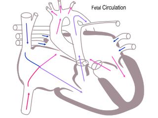
Human Fetal Circulation Mahdia Shaker
Overview of Conversion • Umbilical cord is clamped • Loose placenta • Closure of ductus venosus • Blood is transported to liver and portal system • Increased systemic resistance • Pressure in right atrium decreased • Change from right to left shunting to left to right blood flow • Increased O2 levels in pulmonary circulation • Closure of the ductus arteriosus Mahdia Shaker
Loss of placenta also leads to: • First breath • Lungs expand and fluid is expelled • Decreased pulmonary resistance • Increased pressure in left atrium • Closure of foramen ovale • Increased systemic resistance • Pressure in right atrium decreased • Change from right to left shunting to left to right blood flow • Increased O2 levels in pulmonary circulation • Closure of the ductusarteriosus Mahdia Shaker
Fetal vs. Infant Circulation Fetal • Low pressure system • Right to left shunting • Lungs non-functional • Increased pulmonary resistance • Decreased systemic resistance Infant • High pressure system • Left to right blood flow • Lungs functional • Decreased pulmonary resistance • Increased systemic resistance
Shunts Occur when the foramen ovale or ductusarteriosus remains open, placing a strain on the heart. • In patent foramen ovale (atrial septum defect), the blood flows from left atrium to right atrium (left-to-right shunt) lead to right ventricle (volume overload) , lungs and then left atrium. Unfortunately in about 1 of every 5500 babies the ductusarteriosus never closes. • In patent ductusarteriosus, the blood flows from aorta to pulmonary artery (= left-to-right shunt) leads to lungs (pressure overload) and then aorta. Mahdia Shaker
Flow Chart of Fetal Circulation Mahdia Shaker