Fetal Circulation
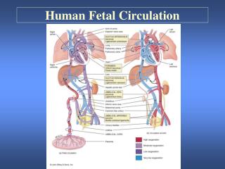
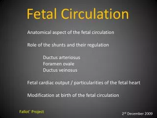
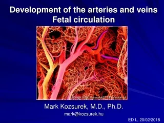
Fetal Circulation. Fetal Circulation. By the third month of development, all major blood vessels are present and functioning. Fetus must have blood flow to placenta. Resistance to blood flow is high in lungs. Umbilical Circulation.

Fetal Circulation
E N D
Presentation Transcript
Fetal Circulation • By the third month of development, all major blood vessels are present and functioning. • Fetus must have blood flow to placenta. • Resistance to blood flow is high in lungs.
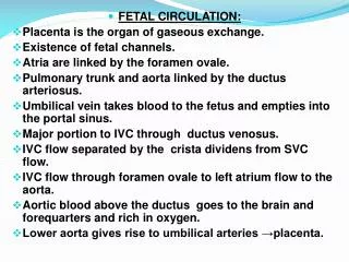
Umbilical Circulation • Pair of umbilical arteries carry deoxygenated blood & wastes to placenta. • Umbilical vein carries oxygenated blood and nutrients from the placenta.
The Placenta • Facilitates gas and nutrient exchange between maternal and fetal blood. • The blood itself does not mix.
Umbilical vein to portal circulation • Some blood from the umbilical vein enters the portal circulation allowing the liver to process nutrients. • The majority of the blood enters the ductus venosus, a shunt which bypasses the liver and puts blood into the hepatic veins.
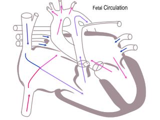
foramen ovale • Blood is shunted from right atrium to left atrium, skipping the lungs. • More than one-third of blood takes this route. • Is a valve with two flaps that prevent back-flow.
ductus arteriousus • The blood pumped from the right ventricle enters the pulmonary trunk. • Most of this blood is shunted into the aortic arch through the ductus arteriousus.
What happens at birth? • The change from fetal to postnatal circulation happens very quickly. • Changes are initiated by baby’s first breath.
Problem with persistence of fetal circulation • Patent (open) ductus arteriosus and patent foramen ovale each characterize about 8% of congenital heart defects. • Both cause a mixing of oxygen-rich and oxygen-poor blood; blood reaching tissues not fully oxygenated. Can cause cyanosis. • Surgical correction now available, ideally completed around age two. • Many of these defects go undetected until child is at least school age.