Download
1 / 53
530 likes | 573 Views
Explore causes, classifications, and prevention strategies for bacteremia and endocarditis. Learn about microbiologic etiology and pathogenesis factors involved in these infections. Stay informed on current recommendations.

E N D
Bacteremia and Endocarditis September 26th, 2005
2 sets, >15 minutes apart Now continuous monitoring No entry of bacteria for monitoring lysis-centrifugation system (Isolator) used for either routine bacteria, fungi, mycobacteria, or fastidious organisms such as Bartonella or Brucella Labor intensive More contamination Technique 70% alcohol, followed by tincture of iodine (1 minute) or povidone iodine (2 minutes). septum of the culture bottle or tube need only be wiped with 70% alcohol transported to the laboratory promptly volume of blood cultured and the number of sets drawn are particularly important current recommendations for adults are to draw at least two separate blood cultures totaling 30 to 40 ml of blood. Separate venipunctures should be performed to help interpret cultures that contain skin flora Blood Cultures
Classification of Bacteremia • Community-acquired • Nosocomial • Healthcare-associated Bacteremia • indwelling catheters • HD • receiving other outpatient therapy • Wound care • nursing home residents
Friedman: Ann Intern Med, Volume 137(10).November 19, 2002.791-797
Martin: N Engl J Med, Volume 348(16).April 17, 2003.1546-1554
Infectious Endocarditis (IE) • Traditional risk factor of RHD decreasing, newer factors increasing • Emergence of enterococci and Staph, particularly MRSA and VRE, also VISA and VRSA • Viridans Strep now emerging MDR strains • Subtypes • native value IE • RHD • MVP (10-100 fold increased risk if regurgitant) • Degenerative/inherited valve disorders • prosthetic valve IE • Early (Staph epi, Staph aureus) • Late (Strep, HACEK) • IE in intravenous drug users • Healthcare-associated IE • nosocomial IE
Microbiologic Etiology in 1779 Patients With Definite Endocarditis From: Fowler: JAMA, Volume 293(24).June 22/29, 2005.3012–3021
Trends in Age- and Sex-Adjusted Incidence Rates of Infective Endocarditis Caused by Staphylococcus aureus and Viridans Group Streptococci From 1970 to 2000 in Olmsted County, Minnesota From: Tleyjeh: JAMA, Volume 293(24).June 22/29, 2005.3022–3028
IDU-associated IE • median age 30 and 40 yrs • up to 40% of cases of IE in San Francisco • tricuspid valve > 50% of cases, aortic in 25%, mitral in 20%, mixed right- and left-sided IE unusual. • injections of impure drugs and particulates might produce microtrauma to the tricuspid leaflets, facilitating microbial colonization • 20–40% of IDU with IE have pre-existing cardiac lesions • Bacteria often originate from the skin • streptococci and others also seen • Pseudomonas aeruginosa and fungi may produce severe IE. • mortality of IE higher in patients who have AIDS
Nosocomial IE • The incidence is increasing. • Many patients have other debilitating underlying • < 50% had obvious cardiac predisposing factors • In most circumstances a potential source of bacteremia could be identified, (lines, procedures) • staphylococci and enterococci most common • other organisms-Gram-negative bacteria and fungi. • Right-sided IE is increasingly recognized in association with central venous lines, pulmonary artery catheters and pacemakers. • procedures that produce transient bacteremia represent risk in hospitalized patients, especially when the circulating organism is S. aureus. • mortality of nosocomial IE is greater than 50%.
Culture-negative IE • HACEK • Haemophilus spp. • Actinobacillus actinomycetemcomitans • Cardiobacterium hominis • Eikenella corrodens • Kingella kingae • Nutritionally-deficient Strep spp. • Fastidious GNR
Culture-negative IE • Coxiella burnetii (Q fever) • Brucella • Bartonella quintana • Chlamydia • Tropheryma whippelii
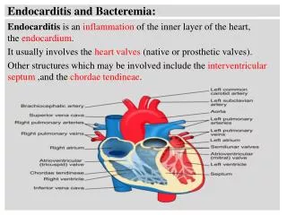
Pathogenesis • Basic lesion is endothelial damage • IE pathogens possess surface ligands that mediate attachment to extracellular matrix proteins of the host (MSCRAMMs) • direct invasion of endothelial cells may also occur • Transient bacteremias occur during chewing, toothbrushing and other normal activities, and from more invasive procedures
Microscopic appearance of a vegetation from a patient suffering mitral valve infective endocarditis due toStreptococcus sanguis. The purple area represents clusters of streptococci packed within a fibrin-platelet meshwork. Professional phagocytes are essentially absent from the lesion.
IE Prophylaxis • Goals: • identification of patients at risk; • determination of the procedures or circumstances that may result in bacteremias; • choice of an appropriate antimicrobial regimen; and • balancing of the known risks against the possible benefits of intervention. • No controlled studies • Regimens used in humans are based upon their proven efficacy in animal models of IE • successful prophylaxis does not require bactericidal antibiotics • Antiseptic mouth rinses applied immediately prior to dental procedures may reduce the incidence or magnitude of bacteremia • Cover most probable pathogens circulating in the blood during a given procedure • oropharyngeal manipulation-streptococci • gastrointestinal or urogenital manipulations, it should be aimed at enterococci (plus results of a preprocedure urine culture) • skin or other infected lesions-staphylococci • If already on antibiotics, choose another class.
Diagnosis • Duke criteria • Classic exam/lab findings • Osler nodes • Splenomegaly • Janeway lesions • Microscopic hematuria • Elevated ESR and CRP • Septic pulmonary emboli (right) • Classic findings may be absent • Repeating TEE 7 to 10 days after an initial "negative" result may be advisable • Posttreatment echocardiography is recommended
Peripheral Stigmata of IE • Osler node=small, raised, tender cutaneous lesion, usually on the pads of fingers or toes (vasculitic) • Janeway lesion=flat, painless small hemorrhages with a slightly nodular appearance that occur on the palms and soles (septic emboli) • Splinter hemorrhages • Petechiae • Roth spots=hemorrhage in the retina with a white center
Surgery • Indication • Severe CHF (reduced mortality with surgery) • Persistent bacteremia • Certain pathogens (fungal, GNR) • Embolic events • Large vegetation on mitral valve highest risk • Valve abscess/dehiscence • Timing • 7-fold higher risk of recurrent IE if valve replaced during active infection
Treatment • If using synergistic agents give them together • Day 1 is first day documented negative blood cultures • postoperative treatment regimen should be one that is recommended for prosthetic valve treatment rather than one that is recommended for native valve treatment • Start over if cultures positive
community-acquired native valve endocarditis in patients who are not intravenous drug users (IDUs). -hemolytic S sanguis, S oralis (mitis), S salivarius, S mutans, and Gemella morbillorum (formerly called S morbillorum). S anginosus group (S intermedius, anginosus, and constellatus) aka S milleri group tends to form abscesses and cause hematogenously disseminated infection (eg, myocardial and visceral abscesses, septic arthritis, vertebral osteomyelitis). S intermedius usually is sensitive to penicillin, but some strains may exhibit variable penicillin resistance. Abiotrophia defectiva and Granulicatella species (G elegans, G adiacens, G paraadiacens, and G balaenopterae; formerly known as nutritionally variant streptococci), have nutritional deficiencies that hinder their growth Gemella (morbillorum, bergeriae, sanguinis, and hemolysans) share some physiological characteristics with nutritionally variant streptococci should be treated with more aggressive combination therapy S bovis expresses the group D antigen, but it can be distinguished from group D Enterococcus by appropriate biochemical tests. should undergo colonoscopy Viridans Strep
Treatment • Native valve, highly susceptible viridans Strep or Strep bovis (MIC <.12) • Aq Pen G x 4 wks • Rocephin x 4 wks • Aq Pen G + gent x 2 wks (synergy) • Rocephin + gent x 2 wks • Vanc x 4 wks • Viridans Group Streptococci and S bovis With Penicillin MIC >0.12 to 0.5 µg/ml • Aq pen or Rocephin x 4 wks + gent 1st 2 wks (single daily dose) • vancomycin
Treatment • A defectiva, Granulicatella species, and Gemella species and a microorganism with an MIC to penicillin >0.5 µg/mL should be treated with a regimen that is recommended for enterococcal endocarditis • prosthetic valves should receive 6 weeks of therapy with penicillin or ceftriaxone with or without gentamicin for the first 2 weeks • S pneumoniae, S pyogenes, and Groups B, C, and G Streptococci • highly penicillin-susceptible S pneumoniae should receive 4 weeks of antimicrobial therapy with penicillin, cefazolin, or ceftriaxone • High-dose penicillin or a third-generation cephalosporin can be used in patients with penicillin-resistant infection and without meningitis • If the isolate is resistant (MIC 2 µg/mL) to cefotaxime, then the addition of vancomycin and rifampin should be considered. • Consider gentamicin for at least the first 2 weeks of a 4- to 6-week course of antimicrobial therapy for group B, C, and G strep (relatively pen res) • Aq pen G for Gp A
Usually associated with PVE Occasionally native valve, usually damaged More indolent than Staph aureus Staph lugdinensis more virulent Coagulase-negative Staph (CoNS)
Nosocomial bacteremia previously thought to be lower risk for endocarditis health care–associated infection was the single most common form of S aureus IE health care–associated IE is distinguished by a relative infrequency of classic clinical stigmata of IE S aureus bacteremia associated with health care has increased among hospitalized patients and among those receiving outpatient medical therapy MRSA in both hospital and community increased dramatically implanted medical devices prosthetic heart valves grafts hemodialysis catheters pacemakers Staph aureus endocarditis
Endocarditis • Factors associated with Staph aureus SBE: • Native valve • Hemodialysis • Invasive procedures • Other chronic disease • Multiple pulmonary emboli • Intravascular device source • Tricuspid • Healthcare-associated • IDU-associated • Persistent bacteremia, emboli requiring surgery • Complications including stroke, other emboli, death • Factors associated with non-Staph aureus SBE • Aortic valve • Prosthetic valve • Congenital heart disease • Dental work • Symptoms >1 month International Collaboration on Endocarditis-Prospective Cohort Study from June 2000 to December 2003.
Figure. In-Hospital Mortality Rates Among Patients With Health Care–Associated Staphylococcus aureus Endocarditis. Includes both nosocomial and nonnosocomial health care–associated infections, community-acquired injection drug use–associated S aureus endocarditis, and community-acquired noninjection drug use–associated S aureus endocarditis by geographic region. From: Fowler: JAMA, Volume 293(24).June 22/29, 2005.3012–3021
Table 4. Clinical Characteristics and Outcomes of 424 Prospectively Identified Patients With Definite Endocarditis Due to Methicillin-Susceptible and Methicillin-Resistant Staphylococcus aureus*From: Fowler: JAMA, Volume 293(24).June 22/29, 2005.3012–3021
Treatment • Staph aureus IDU (right) • parenteral ß-lactam +/- gent x 2 wks (uncomplicated) • Oral also effective (Cipro + rifampin x 4 wks) • Vancomycin requires 4 wks • Staph aureus non-IDU • Beta lactam x 4-6wks, gent 1st few days if fulminant • Faster clearance of bacteremia • Not better mortality • Vanc only if anaphylactoid history to PEN • Clinda not recommended, high relapse rate • Consider desensitizing or daptomycin. • PVE add gentamicin for 2 wks, plus rifampin full 6 wks
Treatment • MRSA • Vancomycin (same gent caveats, plus possible increased ototox) • Linezolid • Synercid • Daptomycin • PVE add gentamicin for 2 wks, plus rifampin full 6 wks • CoNS • Assume meth-resistant • Vancomycin for 6 wks • PVE add gentamicin for 2 wks, plus rifampin full 6 wks
Enterococci • Group D • test MICs to penicillin and vancomycin and for high-level resistance to gentamicin and streptomycin • trough antibiotic concentration in serum must be maintained above the MIC. • relatively resistant to penicillin, ampicillin, and vancomycin. • requires the synergistic action of penicillin, ampicillin, or vancomycin in combination with either gentamicin or streptomycin. • relatively impermeable to aminoglycosides. • cell wall–active agents raise the permeability of the enterococcal cell so that a bactericidal effect can be achieved • If resistant to high concentrations of an aminoglycoside (500 µg/mL of gentamicin or 1000 µg/mL of streptomycin), then the combination of an aminoglycoside with the cell wall–active agent will not result in bactericidal activity, nor will it predictably produce a microbiological cure. • <3 months’ duration of symptoms 4 weeks; >3 months’ duration of symptoms 6 weeks • PVE 6 wks
Enterococci • Beta lactam resistant-vancomycin • VRE • vancomycin-resistant E faecalis and E gallinarum/casseliflavus usually are penicillin susceptible • Linezolid • Daptomycin • Synercid (faecium only)
fastidious Gram-negative bacilli (grow slowly ) Haemophilus parainfluenzae, H aphrophilus, H paraphrophilus, H influenzae, Actinobacillus actinomycetemcomitans, Cardiobacterium hominis, Eikenella corrodens, Kingella kingae, and K denitrificans 5%-10% of native valve community-acquired IE in non IDUs hold blood cultures for 2 wks in patients suspected of having IE. ß-lactamase–producing strains increasing should be considered ampicillin resistant susceptible to ceftriaxone, ampicillin-sulbactam, and fluoroquinolones. limited published clinical data duration of therapy for native valve infection should be 4 weeks prosthetic valve 6 weeks bacteremia caused by HACEK is highly suggestive of endocarditis HACEK
IDU, prosthetic valve, and cirrhosis are risk factors Enterobacteriaceae Salmonella species have an affinity for abnormal cardiac valves Valvular perforation, atrial thrombi, myocarditis, and pericarditis are common Salmonellae also may produce endarteritis in aneurysms of major vessels. Other Enterobacteriaceae, including E coli and Serratia marcescens, may rarely cause endocarditis S marcescens endocarditis typically develops in IDUs. Left-sided disease, large vegetations, and involvement of normal valves mortality rates are 70%. Cardiac surgery is a cornerstone of treatment Combinations of pens/cephs and aminoglycosides have been shown to be synergistic E. coli or Proteus mirabilis, a combination of either ampicillin or penicillin or a broad-spectrum ceph + aminoglycoside, usually gent Endovascular Salmonella infections also may respond to third-generation cephalosporins. combination of a third-generation cephalosporin and an aminoglycoside (either gentamicin or amikacin) is recommended for Klebsiella endocarditis. Other Gram-negatives
Nearly all IDUs Associated with tripelennamine and pentazocine ("T’s and blues") mean age 30 years affects normal valves Major embolic phenomena, inability to sterilize valves, neurol complications (53%), ring and annular abscesses, splenic abscesses, bacteremic relapses, and rapidly progressive CHF are common. many authorities recommend early surgery for left-sided Pseudomonas endocarditis High-dose regimens of antipseudomonal penicillins combined with aminoglycosides are used minimum 6 weeks Medical therapy works in P aeruginosa IE involving the right side of the heart in 50% to 75% partial tricuspid valvulectomy or "vegetectomy“ if failure quinolones (in combination with an aminoglycoside) appear promising, based on favorable results in animal models and humans, but development of stepwise resistance during therapy may limit the efficacy ceftazidime-tobramycin is preferred over aztreonam-tobramycin 7 cases of P aeruginosa endocarditis have been successfully treated with imipenem plus an aminoglycoside potential for the development of resistance exists with any of these regimens. Pseudomonas
Culture-negative IE • Why? • Antibiotics prior • Cover your bases i.e. Staph, Strep, enterococcus • Fastidious organism (Rocephin + gent +doxy) • Bartonella • Brucella • Q fever • Whipple’s • Chlamydia • Not infectious • Marantic • Autoimmune • Neoplastic
often a complication of medical and surgical advances usually have multiple predisposing conditions (cardiovascular devices, prosthetic cardiac valves and central venous catheters) mortality rates for fungal endocarditis are very high. survival rate for patients with mold-related endocarditis is <20%. Candida and Aspergillus species account for the large majority Historically, is an indication for surgical replacement of an infected valve. amphotericin B, a fungicidal agent, is the drug of choice antifungal therapy usually is given for 6 weeks. long-term (lifelong) suppressive therapy with an oral azole Fungal IE
Predisposing Conditions in FEFrom: Pierrotti: Chest, Volume 122(1).July 2002.302-310