1 / 0

Animal Nutrition

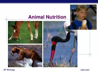
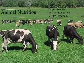
Animal Nutrition. Chapter 41. Evolutionary Development. Four Types of Ingestion/ Feeding Mechanisms Substrate Feeders Live on or in food and eat their way through Ex.: Caterpillars Fluid Feeders Suck nutrient-rich fluid from a living organism Ex.: Mosquitoes, Hummingbirds

joben
Download Presentation

Animal Nutrition

An Image/Link below is provided (as is) to download presentation Download Policy: Content on the Website is provided to you AS IS for your information and personal use and may not be sold / licensed / shared on other websites without getting consent from its author. Content is provided to you AS IS for your information and personal use only. Download presentation by click this link. While downloading, if for some reason you are not able to download a presentation, the publisher may have deleted the file from their server. During download, if you can't get a presentation, the file might be deleted by the publisher.

E N D

Presentation Transcript


  1. Animal Nutrition

    Chapter 41
  2. Evolutionary Development Four Types of Ingestion/ Feeding Mechanisms Substrate Feeders Live on or in food and eat their way through Ex.: Caterpillars Fluid Feeders Suck nutrient-rich fluid from a living organism Ex.: Mosquitoes, Hummingbirds Suspension Feeders Sift small food particles from the water Ex.: Whales, clams, oysters Bulk Feeders Most animals Eat large pieces of food with a variety of adaptations
  3. Evolutionary Development Digestive Compartments- reduce the risk of self-digestion by hydrolytic enzymes Intracellular digestion Occurs in vacuoles, organelles in individual cells Simplest digestive compartments Food vacuoles fuse with lysosomes to digest food Extracellular digestion Occurs in larger compartments continuous with the outside of the body Allows animals to ingest larger prey
  4. Evolutionary development-An animal’s diet can bring about different adaptations to the basic, common digestive system Stomach and Intestinal Adaptations Carnivores, who often have a longer time between meals, have bigger, expandable stomachs so that they can eat larger quantities at one time. Herbivores and Omnivores generally have longer alimentary canals than carnivores (relative to body size). Plant matter is harder to digest because it contains cell walls, so a longer digestive tract allows more time for adequate digestion and absorption. Dental Adaptations Carnivores, animals that preferably eat meat, have sharp and pointed incisors and canines to rip and tear flesh and muscle of prey. The premolars and molars crush and grind food. Herbivores, animals that preferably eat plants, have wide, broad teeth that grind food. The incisors and canines aren’t as sharp as those of carnivores, instead they are better suited for biting off vegetation or completely absent. Omnivores, animals that eat both plants and other animals, have unspecialized dentition due to their diverse diets. Fangs are modified teeth that inject venom. Fangs can either be hollow to fill with venom or grooved to drip poison. Snakes jaws lower jaws are very loosely hinges so that they can open their mouths wide to swallow larger prey.
  5. More evolutionary adaptations Symbiotic Adaptations Most of the nutrients and chemical energy from plant food is found in the cell walls. Herbivores, however, don’t have enzymes that can break down cellulose. So, herbivores house symbiotic bacteria and protists within their alimentary canals that can digest cellulose into smaller nutrients that the animal can use. Examples in microorganism locations In Hoatzins, South American birds, symbionts live in the birds’ crops. Many mammals, like horses, have microorganisms living in their cecums. Rabbits and rodents have helpful bacteria in their large intestines. Ruminants, including deer, cows, and sheep, have the most elaborate housing system for symbionts. Ruminants have a four-chambered stomach that houses many microorganisms *Major Theme of Biology: “Relationship of Structure to Function”
  6. Extracellular digestion Types Gastro vascular Cavity – simple digestive sac with one opening in cnidarians and flatworms Responsible for both digestion and distribution of nutrients Gastro dermis tissue secretes enzymes to break down prey and nutritive muscular cells engulf molecules for intracellular digestion Complete Digestive Tract- “Alimentary Canal” Digestive tube with two openings, a mouth and an anus Specialized regions of tract allow for stepwise digestion
  7. Necessary for… Food provides an animal with fuel to produce ATP and with raw materials growth and survival Undernourishment-diet is deficient in calories Malnourishment- diet is deficient in any essential nutrient Carbon skeletons from an animal’s food are used to build the animal’s own structures Essential nutrients cannot be made from any raw materials by an animal, and therefore have to come from food Vitamins Minerals Essential Fatty Acids Essential Amino Acids *Major Theme of Biology: “Interdependence in Nature”
  8. Essential Amino Acids 8 of the 20 amino acids needed by humans have to come from food. Protein deficiency, a type of malnourishment, occurs when an animal’s diet is missing sufficient amounts of an essential amino acid. Kwashiorkor- a severe protein deficiency that usually affects children living in poverty Symptoms include swelling (edema), a protruding stomach, and extreme tiredness and muscle fatigue (“PubMedHealth”)
  9. Vitamins Organic molecules required in relatively small amounts 13 essential vitamins (for humans) have been identified Water-Soluble: Vitamins B1, B2, B6, B12, C, Niacin, Pantothenic acid, Folic acid, and Biotin Coenzymes in many different metabolic processes Fat-Soluble Vitamins A and E act as antioxidants and prevent cell membrane damage Vitamin D promotes bone growth and calcium use and absorption Vitamin K is essential to blood clotting
  10. Minerals Simple inorganic nutrients required in small amounts Important minerals for humans Calcium – blood clotting, nerve formation, and bone growth Iron- in hemoglobin and in electron carriers, also an enzyme cofactor Phosphorous- helps in nucleotide synthesis Sulfur, Potassium, Chlorine, Sodium, Magnesium, Fluorine, Zinc, Copper, Manganese, Iodine, Cobalt, Selenium, Chromium, and Molybdenum
  11. Human structure and function http://www.youtube.com/watch?v=08VyJOEcDos Four Stages of Animal Nutrition Ingestion-the physical act of eating food Digestion-the breakdown of food into small molecules the body can absorb via enzymatic hydrolysis, adding water to break the bonds of food macromolecules Absorption-the animal’s cells take up smaller molecules like simple sugars and amino acids broken down from the food Elimination- waste products of undigested materials pass out of the animal’s body.
  12. Oral cavity The mouth Teeth and tongue facilitate mechanical digestion. Salivary glandssecrete saliva containing mucus to protect mouth cells salivary amylase to hydrolyze starch and glycogen, beginning chemical digestion Tongue forms food into a ball called a “bolus” and moves it to the pharynx http://training.seer.cancer.gov/head-neck/anatomy/overview.html
  13. Pharynx and esophagus Pharynx Esophagus The bolus moves down the esophagus, from the mouth to the stomach, thanks to peristalsis Peristalsis- rhythmic, repeated involuntary muscle contractions of the alimentary canal The throat Opens to both the windpipe and the esophagus A cartilage flap called the epiglottis closes over the windpipe when swallowing, to prevent food or water from entering the airway.
  14. stomach responsible for food storage and primary digestion Accordion-like folds and an elastic wall allow large food storage Cardiac Sphincter- located at the top of the stomach to keep food from moving back up into the esophagus Pyloric Sphincter- located at the bottom of the stomach to keep food in the stomach long enough for sufficient digestion All mammals have rennin in their stomachs to digest the proteins in milk http://www.naturalhealthschool.com/6_2.html
  15. Gastric Juice in the stomach: Has a pH of 2 to dissolve the ECM of food and to kill bacteria Contains pepsin, which digests proteins in food Chief cells secrete pepsinogen, the inactive form Pepsinogen in activated by hydrochloric acid, which is secreted by parietal cells Mucus cells secrete mucus to protect stomach cells from self digestion Acid Chime, the nutrient-rich broth of digested food, moves into the small intestine. Stomach http://www.tutorvista.com/content/biology/biology-ii/nutrition/human-digestive-system.php
  16. Small intestine responsible for most of enzymatic hydrolysis of food and most absorption of nutrients into blood stream Duodenum- first 25 cm in which acid chime mixes with digestive juices from pancreas, liver, and gallbladder. Most digestion is done here. Bile- digestive juice produced by liver and stored in gallbladder. Bile digests fats. Peptidases break down proteins Nucleases hydrolyze nucleic acids Lipases break down fats
  17. Small Intestine The remaining lengths of the small intestine absorb nutrients and water into the bloodstream. Villi-projections on the surface of the small intestine that allow for more surface area, helping absorption Capillaries within the villi absorb nutrients, including amino acids and vitamins Lacteals-lymph vessels inside villi that attract chylomicron Chylomicrons- small globules of fats, cholesterol, and coated with proteins Hepatic portal vein- takes blood with absorbed nutrients from small intestine to the liver
  18. Large intestine Cecum: The large intestine connects to the small at a T-shaped junction. The cecum is one arm of that “T”. The appendix is an appendage of the human cecum. It is the first section of the large intestine and collects waste material from the small intestine Colon: The rest of the large intestine is split into different “colon” sections The waste is solidified into feces as it makes its way through the colon Feces is then stored in the rectum until it can be eliminated http://en.wikipedia.org/wiki/File:Intestine-diagram.svg
  19. Interdependence with other body systems Circulatory System Nutrients digested throughout the digestive system are absorbed into the bloodstream. They are then carried throughout the body via the circulatory system. Lacteals, located in the villi of the small intestine, are part of the lymphatic/ circulatory system. Lacteals attract chylomicrons which bring chyle, a type of lymph, into the circulatory system.
  20. Interdependence with other body systems Endocrine System Excess calories are stored as glycogen and as fat. When an animal has to use these stores for energy, its metabolism is regulated by negative feedback. After eating, the level of glucose in the blood rises. The pancreas gland secretes a hormone insulin into the blood stream. Insulin helps transport glucose and stimulates the storage of glucose as glycogen by the muscles and liver. Blood glucose level drops. When the level of glucose in the blood stream drops, the pancreas secretes another hormone called glucagon. Glucagon has the opposite effect of insulin. Glycogen breakdown in the liver and the release of more glucose into the blood stream are stimulated by glucagon. As a result, the amount of glucose in the blood stream increases. Endocrine System Hormones are released by the stomach and duodenum to control digestion. Cholecystokinin is released when amino and/or fatty acids are present in the duodenum. Cholecystokinin stimulates the release of enzymes from the pancreas and bile from the gallbladder Enterogastroneslows digestion when fatty acid chime enters the duodenum. The duodenum secretes enterogastrone to inhibit peristalsis and acid secretion Secretin is secreted by the duodenum to activate pancreas release of sodium bicarbonate to neutralize acid chime Gastrin starts the production of gastric juices *Major Theme of Biology: Regulation
  21. Diseases of the Digestive system Diverticulitis – the inflammation or infection of small pouches the inner lining of the intestines. Causes Possibly a low-fiber diet may cause Diverticulosis, the formation of small pouches. Eating too many processed foods without enough fiber is harmful to the digestive system A low-fiber diet may cause hard stool and constipation, which put way too much pressure on the intestines. This increase in pressure may cause the small pouches to form in the inner lining. If feces gets caught in any of these pouches, the pouch becomes inflamed or infected.
  22. diverticulitis Symptoms People rarely exhibit symptoms of diverticulosis, except bloating of the lower abdomen. The symptoms of diverticulitis are more severe. Tenderness of the lower left abdomen Nausea and vomiting Loss of appetite Bloating and gas Fever and Chills
  23. Diverticulitis Treatment and Medication Severe cases of diverticulitis require hospitalization, but most cases can be treated at home Possible treatments include antibiotics, a liquid-only diet for a few days, or pain medications prescribed by a doctor. Increasing the amount of fiber and decreasing the amount of processed foods in a diet can help avoid attacks of diverticulitis. The pouches of the inner lining of the intestines from diverticulosis are manageable, but will not go away.
  24. Appendicitis Appendicitis- swelling of the appendix Causes Something blocks the appendix, maybe feces, a tumor, or another object Symptoms Early Symptoms: The area around the belly button will start to hurt. The pain may vary in intensity. Loss of appetite, nausea, and a fever If swelling of the appendix continues, The pain will migrate to the lower, right-hand side of the abdomen above the appendix. Constipation, diarrhea, chills, fever, shaking, nausea, and vomiting If the appendix ruptures, pain will subside for a little bit before intensifying as the lining of the intestinal cavity becomes inflamed and infected.
  25. Appendicitis Treatment and Medications If the doctor does not see any complications and the appendix has not yet ruptured, they will remove the appendix. This surgery is called an appendectomy and the recovery time is relatively short. Appendicitis is very common in the United States. It often results in emergency surgery because it is incredibly hard to detect early on, especially in kids, the elderly, and women.
  26. Citations “Appendicitis." PubMedHealth. A.D.A.M. Inc., 22 Jul 2011. Web. 13 Apr 2012. Campbell, Neil A., and Jane B. Reece. AP Edition Biology. 7th ed. San Francisco: Pearson Education Inc., 2005. Print. “Diverticulitis." PubMedHealth. A.D.A.M. Inc., 31 Jan 2012. Web. 13 Apr 2012. Goldberg, Deborah T. Barron's AP Biology. 3rd ed. Barron's Educational Series Inc., 2010. Print. "Hepatic Portal Vein." The American Heritage Medical Dictionary. Houghton Mifflin Company, 2007. Web. 22 Apr 2012. <http://medical-dictionary.thefreedictionary.com>. "Kwashiorkor." PubMedHealth. A.D.A.M. Inc., 01 Feb 2012. Web. 13 Apr 2012. Meyers, Donna. "Cecum." Colon Cancer. About.com, 08 Apr 2007. Web. 22 Apr 2012. "What Is a Lacteal?." wiseGEEK. wiseGEEK, n.d. Web. 22 Apr 2012.
More Related