Download
1 / 51
510 likes | 549 Views
Explore the groundbreaking experiments of Griffith, Avery, and Hershey-Chase that established DNA as the hereditary material. Learn about the structure of DNA and how it replicates.

E N D
9.1 The Griffith Experiment • Mendel’s work left a key question unanswered: • What is a gene? • The work of Sutton and Morgan established that genes reside on chromosomes • But chromosomes contain proteins and DNA • So which one is the hereditary material • Several experiments ultimately revealed the nature of the genetic material
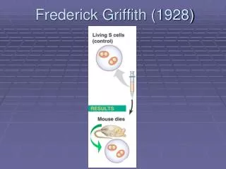
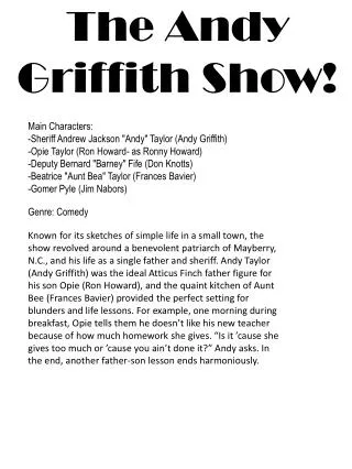
9.1 The Griffith Experiment • In 1928, Frederick Griffith discovered transformation while working on Streptococcus pneumoniae • The bacterium exists in two strains • S • Forms smooth colonies in a culture dish • Cells produce a polysaccharide coat and can cause disease • R • Forms rough colonies in a culture dish • Cells do not produce a polysaccharide coat and are therefore harmless
Fig. 9.1 How Griffith discovered transformation Thus, the dead S bacteria somehow “transformed” the live R bacteria into live S bacteria
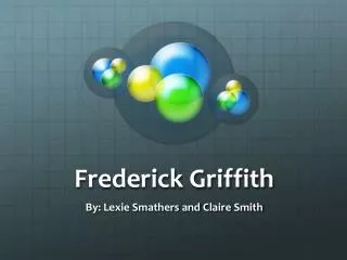
Two key experiments that demonstrated conclusively that DNA, and not protein, is the hereditary material Oswald Avery and his coworkers Colin MacLeod and Maclyn McCarty published their results in 1944 Alfred Hershey and Martha Chase published their results in 1952 9.2 The Avery and Hershey-ChaseExperiments
The Avery Experiments • Avery and his colleagues prepared the same mixture of dead S and live R bacteria as Griffith did • They then subjected it to various experiments • All of the experiments revealed that the properties of the transforming principle resembled those of DNA • 1. Same chemistry and physical properties as DNA • 2. Not affected by lipid and protein extraction • 3. Not destroyed by protein- or RNA-digesting enzymes • 4. Destroyed by DNA-digesting enzymes
The Hershey-Chase Experiment • Viruses that infect bacteria have a simple structure • DNA core surrounded by a protein coat • Hershey and Chase used two different radioactive isotopes to label the protein and DNA • Incubation of the labeled viruses with host bacteria revealed that only the DNA entered the cell • Therefore, DNA is the genetic material
Fig. 9.2 The Hershey-Chase Experiment Thus, viral DNA directs the production of new viruses
9.3 Discovering the Structure of DNA • DNA is made up of nucleotides • Each nucleotide has a central sugar, a phosphate group and an organic base • The bases are of two main types • Purines – Large bases • Adenine (A) and Guanine (G) • Pyrimidines – Small bases • Cytosine (C) and Thymine (T)
Fig. 9.3 The four nucleotide subunits that make up DNA Nitrogenous base 5-C sugar
Fig. 9.4 Rosalind Franklin (1920-1958) • Purines = Pyrimidines • A = T and C = G • Erwin Chargaff made key DNA observations that became known as Chargaff’s rule Rosalind Franklin’s X-ray diffraction experiments revealed that DNA had the shape of a coiled spring or helix
Fig. 9.4 James Watson (1928- ) Francis Crick (1916-2004) • They came to their conclusion using Tinkertoy models and the research of Chargaff and Franklin • In 1953, James Watson and Francis Crick deduced that DNA was a double helix
Fig. 9.4 The DNA double helix The two possiblebasepairs Dimensions suggested by X-ray diffraction
ATACGCAT If the sequence on one strand is TATGCGTA The other’s sequence must be 9.4 How the DNA Molecule Replicates • The two DNA strands are held together by weak hydrogen bonds between complementary base pairs • A and T • C and G • Each chain is a complementary mirror image of the other • So either can be used as template to reconstruct the other
Fig. 9.5 • There are 3 possible methods for DNA replication Daughter DNAs contain one old and one new strand Old and new DNA are dispersed in daughter molecules Original DNA molecule is preserved
Fig. 9.6 • These three mechanisms were tested in 1958 by Matthew Meselson and Franklin Stahl
How DNA Copies Itself • The process of DNA replication can be summarized as such • The enzyme helicase first unwinds the double helix • The enzyme primase puts down a short piece of RNA termed the primer • DNA polymerase reads along each naked single strand adding the complementary nucleotide
Template strand New strand Template strand New strand 3’ HO HO 3’ 5’ 5’ C C P P G G O O Sugar- phosphate backbone O O P P T T A A P P O O O O P P T T A A P P DNA polymerase O O O O P P C C G G P P O O O O P P A 3’ A T OH P P P O O O Pyrophosphate T P P P P P O A A 3’ OH O O OH P P 5’ 5’ Fig. 9.7 How nucleotides are added in DNA replication
Fig. 9.8 • DNA polymerase can only build a strand of DNA in one direction • The leading strand is made continuously from one primer • The lagging strand is assembled in segments created from many primers
Fig. 9.9 RNA primers are removed and replaced with DNA Ligase joins the ends of newly-synthesized DNA Mechanisms exist for DNA proofreading and repair
DNA RNA Protein 9.5 Transcription • The path of genetic information is often called the central dogma • A cell uses three kinds of RNA to make proteins • Messenger RNA (mRNA) • Transfer RNA (tRNA) • Ribosomal RNA (rRNA)
Fig. 9.10 9.5 Transcription • Gene expression is the use of information in DNA to direct the production of proteins • It occurs in two stages
Fig. 9.11 9.5 Transcription • The transcriber is RNA polymerase • It binds to one DNA strand at a site called the promoter • It then moves along the DNA pairing complementary nucleotides • It disengages at a stop signal
9.6 Translation • Translation converts the order of the nucleotides of a gene into the order of amino acids in a protein • The rules that govern translation are called the genetic code • mRNAs are the “blueprint” copies of nuclear genes • mRNAs are “read” by a ribosome in three-nucleotide units, termed codons • Each three-nucleotide sequence codes for an amino acid or stop signal
Fig. 9.12 • The genetic code is (almost) universal • Only a few exceptions have been found
Fig. 9.13 Ribosomes • The protein-making factories of cells • They use mRNA to direct the assembly of a protein A ribosome is made up of two subunits • Each of which is composed of proteins and rRNA Sites play key roles in translation
Fig. 9.14 Transfer RNA Hydrogen bonding causes hairpin loops • tRNAs bring amino acids to the ribosome • They have two business ends • Anticodon which is complementary to the codon on mRNA • 3’–OH end to which the amino acid attaches 3-D shape
Fig. 9.16 Making the Protein • mRNA binds to the small ribosomal subunit • The large subunit joins the complex, forming the complete ribosome • mRNA threads through the ribosome producing the polypeptide
Fig. 9.15 How translation works • The process continues until a stop codon enters the A site • The ribosome complex falls apart and the protein is released
9.7 Architecture of the Gene • In eukaryotes, genes are fragmented • They are composed of • Exons – Sequences that code for amino acids • Introns – Sequences that don’t • Eukaryotic cells transcribe the entire gene, producing a primary RNA transcript • This transcript is then heavily processed to produce the mature mRNA transcript • This leaves the nucleus for the cytoplasm
Fig. 9.17 Processing eukaryotic mRNA Protect from degradation and facilitate translation • Different combinations of exons can generate different polypeptides via alternative splicing
7. Phosphorylation or other chemical modifications can alter the activity of a protein after it is translated. 6. The polypeptide chain grows until the protetin is completed. Amino acid Completed polypeptide tRNA 5’ Ribosome moves toward 3’ end Cytoplasm Ribosome 5. tRNAs bring their amino acids in at the A site of the ribosome. Peptide bonds form between amino acids at the P site, and tRNAs exit the ribosome from the E site. 4. tRNA molecules become attached to specific amino acids with the help of activating enzymes. Amino acids are brought to the ribosome in the order dictated by the mRNA. DNA Nuclear membrane 3’ 3’ RNA polymerase Cap 5’ Small ribosomal subunit 5’ 1. In the cell nucleus, RNA polymerase transcribes RNA from DNA Primary RNA transcript 3’ Nuclear pore 5’ 5’ Large ribosomal subunit 3’ Cap Exons Poly-A tail mRNA Poly-A tail Introns mRNA 3. mRNA is transported out of the nucleus. In the cytoplasm, ribosomal subunits bind to the mRNA 3’ 2. Introns are excised from the RNA transcript, and the remaining exons are spliced together, producing mRNA Fig. 9.18 How protein synthesis works in eukaryotes
9.7 Architecture of the Gene • Most eukaryotic genes exist in multiple copies • Clusters of almost identical sequences called multigene families • As few as three and as many as several hundred genes • Transposable sequences or transposons are DNA sequences that can move about in the genome • They are repeated thousands of times, scattered randomly about the chromosomes
9.8 Turning Genes Off and On • Genes are typically controlled at the level of transcription • In prokaryotes, proteins either block or allow the RNA polymerase access to the promoter • Repressors block the promoter • Activators make the promoter more accessible • Most genes are turned off except when needed
The lac Operon • An operon is a segment of DNA that contains a cluster of genes that are transcribed as a unit • The lac operon contains • Three structural genes • Encode enzymes involved in lactose metabolism • Two adjacent DNA elements • Promoter • Site where RNA polymerase binds • Operator • Site where the lac repressor binds
Fig. 9.19 The lac Operon • In the absence of lactose, the lac repressor binds to the operator • RNA polymerase cannot access the promoter • Therefore, the lac operon is shut down
The lac Operon • In the presence of lactose, a metabolite of lactose called allolactose binds to the repressor • This induces a change in the shape of the repressor which makes it fall off the operator • RNA polymerase can now bind to the promoter • Transcription of the lac operon is ON
The lac Operon • What if the cell encounters lactose, and it already has glucose? • The bacterial cell actually prefers glucose! • The lac operon is also regulated by an activator • The activator is a protein called CAP • It binds to the CAP-binding site and gives the RNA polymerase more access to the promoter • However, a “low glucose” signal molecule has to bind to CAP before CAP can bind to the DNA
Fig. 9.21 Enhancers • DNA sequences that make the promoters of genes more accessible to many regulatory proteins at the same time Usually located far away from the gene they regulate Common in eukaryotes; rare in prokaryotes
Bithorax mutant Fig. 9.22 9.9 Mutation • The genetic material can be altered in two ways • Recombination • Change in the positioning of the genetic material • Mutation • Change in the content of the genetic material
9.9 Mutation • Mutation and recombination provide the raw material for evolution • Evolution can be viewed as the selection of particular combinations of alleles from a pool of alternatives • The rate of evolution is ultimately limited by the rate at which these alternatives are generated • Mutations in germ-line tissues can be inherited • Mutations in somatic tissues are not inherited • They can be passed from one cell to all its descendants
Kinds of Mutation • Mutations are caused in one of two ways • Errors in DNA replication • Mispairing of bases by DNA polymerase • Mutagens • Agents that damage DNA
Kinds of Mutation • The sequence of DNA can be altered in one of two main ways • Point mutations • Alteration of one or a few bases • Base substitutions, insertion or deletion • Frame-shift mutations • Insertions or deletions that throw off the reading frame
Kinds of Mutation • The position of genes can be altered in one of two main ways • Transposition • Movement of genes from one part of the genome to another • Occurs in both eukaryotes and prokaryotes • Chromosomal rearrangements • Changes in position and/or number of large segments of chromosomes in eukaryotes