Download

1 / 22
260 likes | 1.07k Views
Comparative Anatomy Muscles. Note Set 8 Chapter 10. Muscles. Two muscle groups: Somatic muscles Operate head, trunk, limbs Locomotion and orientation Visceral muscles Operate visceral skeleton Digestion and respiratory movements. Cranial Nerves to Muscles.

E N D
Comparative AnatomyMuscles Note Set 8 Chapter 10
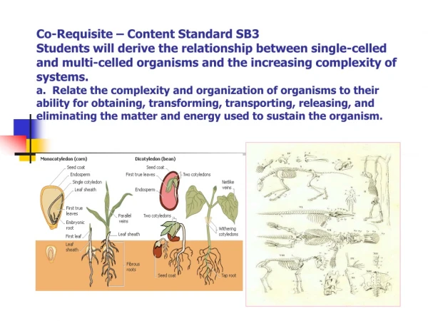
Muscles Two muscle groups: • Somatic muscles • Operate head, trunk, limbs • Locomotion and orientation • Visceral muscles • Operate visceral skeleton • Digestion and respiratory movements
Cranial Nerves to Muscles Figure 10.1: Facial nerves to muscles Figure 10.2: Cranial nerves
Two Muscle Types • Somatic muscles • Skeletal muscle • Striated and voluntary • Visceral muscles • Smooth muscle • Non-striated and involuntary • Exception- branchiomerics (unsegmented)
Skeletal Muscles • Axial • Trunk and tail • Appendicular • Insert on girdles, fins, or limbs • Branchiomerics • Attached to visceral skeleton
Axial • Shark • Epaxial and hypaxial muscles • Body wall muscles • Amphibians • Epaxials above transverse process • Hypaxials along body wall proper • Mammals • Epaxials subdivided • Hypaxials more complex Figure 10.3: Trunk muscles of vertebrates.
Hypaxial and Epaxial Muscles Figure 10.5: Specific epaxial muscles Figure 10.4: Epaxial and hypaxial mucles
Abdominal Muscle Groups in Amniotes • Epaxials • Transversospinalis, longissimus, iliocostalis • Hypaxials • Dorsomedials, laterals, ventrals • Laterals- external oblique, internal oblique, and transverse abdominus • Ventral- rectus abdominus Figure 10.6: Epaxial and hypaxial muscles
Head Region Figures 10.7: Myotomes in the head, neck, and thoracic regions of the embryo. Figure 10.8- Axial muscle origin and innervation in vertebrate embryo.
Head Region • In branchial region, somites are broken down • Ventral slips of postbranchial somites become hypobranchial musculature • Hypobranchial muscles give rise to: • Sternohyoid • Sternothyroid • Omohyoid • Tongue muslces • Geniohyoid • Hyoglossus • Styloglossus • Genioglossus • Lingualis propria
Appendicular Muscles • Extrinsic • Origin on axial skeleton or fascia of trunk • Insert on girdles and limbs • Intrinsic • Origin on girdles or proximal skeletal elements of appendages • Insert on more distal skeletal elements
Intrinsic Muscles Figure 10.9: Intrinsic muscles of pectoral girdle and forelimbs of mammals and their homologues in reptiles.
Branchiomerics • Arises from lateral plate mesoderm • Mandibular (1st) arch • Hyoid arch • Arches IV to VI
Branchiomerics • Mandibular (1st) arch • Intermandibularis- digastic • Adductor mandibulae- masseter, temporalis • Hyoid arch • Sphincter colli • Platysma and mimetics • integumentary muscles • Arches IV to VI • Trapezius, sternomastoid, cleidomastoid Figure 10.10: Branchiomeric muscles of gnathostomes.
Branchiomeric Muscles Figure 10.11: Branchiomeric muscles and their innervations.
Extrinsic Eye Muscles • Six eyeball muscles • 2 obliques • Superior and inferior on anterior portion • 4 rectus • Arise in posterior portion of orbit • Innervated by oculomotor, trochlear, and abducens Figure 10.12: Innervation of eye muscle in embryo.
Extrinsic Eye Muscles Figure 10.13: Dorsal view of extrinsic muscles of the left eyeball. Figure 10.14: Lateral view of extrinsic muscles of eyeball in humans.
Diaphragm • Mammalian muscle structure • Covers lungs and heart in abdominal cavity Figure 10.15: Human diaphragm.
Dermal or Integumentary Muscles • Fish & tailed amphibians- skin is firmly attached to musculature • Sphincter colli- first muscle to move skin • Subdivides down neck- platysma • Extrinsic and intrinsic muscle groups Figure 10.16: Evolution of mammalian facial muscles. Shows sphincter colli (SC) spreading into platysma (P).
Extrinsic Integumentary Muscles • Costocutaneous muscles- allows rectilinear motion (reptiles--snakes) • Panniculus carnosus-sheet surrounds body • Cutaneous maximus- to shake skin (higher mammals) • Patagial muscles- bat wings • Auricularis- moves human ear • Caninus muscle- arises with aggression Intrinsic Integumentary Muscles • Arrectores plumarum (birds) & arrectores pilorum (mammals)- errects hair and feathers
Specialized Muscles • Electric organs • In fish • Modified hypaxial muscles Figure 10.17: Electric eel.
Literature Cited Figure 10.1- http://www.city.ac.uk/optometry/Biolabs/cranial%20nerves/cranial_nerves_lab.htm Figure 10.2- http://mywebpages.comcast.net/epollak/PSY255_pix/PSY255_pix.htm Figure 10.3, 10.8, 10.9, 10.11, 10.12, 10.13, 10.16- Kent, George C. and Robert K. Carr. Comparative Anatomy of the Vertebrates. 9th ed. McGraw-Hill, 2001. Figure 10.4 & 10.5- http://www.mut.ac.th/~vet/Anat-html/muscle/muscle.html Figure 10.6, 10.10- http://people.eku.edu/ritchisong/342notes6.htm Figure 10.7- http://connection.lww.com/products/sadler/imagebank.asp Figure 10.14- http://www.bmb.leeds.ac.uk/illingworth/motors/myosin.htm Figure 10.15- http://whyfiles.org/204endurance_training/2.html Figure 10.17- http://www.aqua.org/animals_electriceel.html