Download

1 / 122
1.22k likes | 1.23k Views
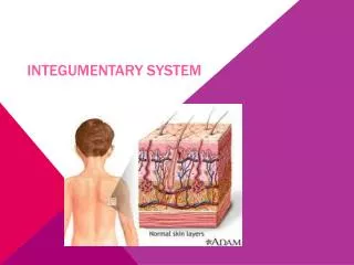
Hair. Nails. Skin. TISSUES (cont.) & THE INTEGUMENTARY SYSTEM Sonya Schuh -Huerta, Ph.D. Human Anatomy. Glands. Ducts carry products of exocrine glands to epithelial surface Include the following diverse glands Mucus-secreting glands Sweat & oil glands Salivary glands

E N D
Hair Nails Skin TISSUES (cont.) & THE INTEGUMENTARY SYSTEM Sonya Schuh-Huerta, Ph.D. Human Anatomy
Glands • Ducts carry products of exocrine glands to epithelial surface • Include the following diverse glands • Mucus-secreting glands • Sweat & oil glands • Salivary glands • Liver & pancreas
Unicellular Exocrine Glands: The Goblet Cell • Goblet cells produce mucin • Mucin + water mucus • Protects & lubricates many internal body surfaces • Goblet cells are a unicellular exocrine gland
Goblet Cells Microvilli Secretory vesicles containing mucin Rough ER Golgi apparatus Nucleus
Multicellular Exocrine Glands • Have 2 basic parts • Epithelium-walled duct • Secretory unit • Classified by structure of duct • Simple • Compound • Categorized by secretory unit • Tubular • Alveolar • Tubuloalveolar
Lateral Surface Features: Cell Junctions • Factors binding epithelial cells together • Adhesion proteins link plasma membranes of adjacent cells • Contours of adjacent cell membranes • Special cell junctions
Lateral Surface Features: Cell Junctions • Tight junctions – close off intercellular space • Found at apical region of most epithelial tissue types • Some proteins in plasma membrane of adjacent cells are fused • Prevent certain molecules from passing between cells of epithelial tissue
Tight Junction Interlocking junctional proteins Intercellular space (a) Tight junctions: Impermeable junctions prevent molecules from passing through the intercellular space.
Lateral Surface Features: Cell Junctions • Adhesive belt junctions (zonula adherens) = anchoring junction • Transmembrane linker proteins attach to actin microfilaments of the cytoskeleton & bind adjacent cells • With tight junctions, these linker proteins form the tight junctional complex around apical lateral borders of epithelial tissues
Lateral Surface Features: Cell Junctions • Desmosomes = main junctions for binding cells together • Scattered along abutting sides of adjacent cells • Cytoplasmic side of each plasma membrane has a plaque • Plaques are joined by linker proteins • Intermediate filaments extend across the cytoplasm and anchor at desmosomes on opposite side of cell • Are common in cardiac muscle & epithelial tissue
Desmosome Intercellular space Plaque Intermediate filament (keratin) Linker glycoproteins (cadherins) (b) Desmosomes: Anchoring junctions bind adjacent cells together and help form an internal tension-reducing network of fibers.
Lateral Surface Features: Cell Junctions • Gap junctions = passageway between 2 adjacent cells • These let small molecules move directly between neighboring cells • Cells are connected by hollow cylinders of protein • Function in intercellular communication!
Gap Junction Intercellular space Channel between cells (connexon) Gap junctions: Communicating junctions allow ions and small molecules to pass from one cell to the next for intercellular communication.
Basal Feature:The Basal Lamina • Non-cellular supporting sheet between the epithelial tissue & the connective tissue deep to it • Consists of proteins secreted by epithelial cells
Basal Feature:The Basal Lamina • Functions: • Acts as a selective filter, determining which molecules from capillaries enter the epithelium • Acts as scaffolding along which regenerating epithelial cells can migrate • Basal lamina & reticular layers of the underlying CT deep to it form the basement membrane
Epithelial Surface Features • Apical surface features • Microvilli = fingerlike extensions of plasma membrane • Abundant in epithelia of small intestine & kidney • Maximize surfacearea across whichsmall moleculesenter or leave • Act as stiff knobsthat resist abrasion Microvillus Actin filaments
Epithelial Surface Features • Apical surface features • Cilia = whiplike, highly motile extensions of apical surface membranes • The apical surface contains a core of 9 pairs of microtubules encircling one middle pair • Axoneme – a set of microtubules • Each pair of microtubules are arranged in a doublet • Microtubules in cilia – arranged similarly to centrioles • Movement of cilia – in coordinated waves
A Cilium Power, or propulsive, stroke Recovery stroke, when cilium is returning to its initial position Outer microtubule doublet The doublets also have attached motor proteins, the dynein arms. Central microtubule Dynein arms The outer microtubule doublets and the two central microtubules are held together by cross-linking proteins and radial spokes. 1 2 3 4 5 6 7 Cross-linking proteins inside outer doublets (b) Phases of ciliary motion Layer of mucus Radial spoke Plasma membrane Cilium Cell surface Basal body (centriole) (a) Structure of a cilium (c) Traveling wave created by the activity of many cilia acting together propels mucus across cell surfaces.
Connective Tissue • Most diverse & abundant tissue • Main classes: • Connective tissue proper • Cartilage • Bone tissue • Blood • Cells separated by a large amount of extracellular matrix • Extracellular matrix is composed of ground substance & fibers • Common embryonic origin – mesenchyme
Mesenchyme (a) Embryonic connective tissue: mesenchyme Description: Embryonic connective tissue; gel-like ground substance containing fibers; star-shaped mesenchymal cells. Mesenchymal cell Ground substance Function: Gives rise to all other connective tissue types. Location: Primarily in embryo. Fibers Photomicrograph: Mesenchymal tissue, an embryonic connective tissue (600); the clear-appearing background is the fluid ground substance of the matrix; notice the fine, sparse fibers.
Structural Elements of Connective Tissue • Connective tissues differ in structural properties • Differences in types of cells • Differences in composition of extracellular matrix • However, connective tissues all share structural elements • Loose areolar connective tissue • Will illustrate connective tissue features
Structural Elements of Connective Tissue • Cells – primary cell type of connective tissue produces matrix • Fibroblasts • Make protein subunits (protein fibers) • Secrete molecules that form the ground substance • Chondroblasts – secrete matrix in cartilage • Osteoblasts – secrete matrix in bone • Blood cells – an exception • Do not produce matrix
Areolar connective tissue: A model connective tissue Extracellular matrix Cell types Ground substance Macrophage Fibers Collagen fiber Elastic fiber Fibroblast Reticular fiber Lymphocyte Fat cell Capillary Mast cell Neutrophil
Structural Elements of Connective Tissue • Fibers – function in support • Collagen fibers – strongest; resist tension • Reticular fibers – bundles of special type of collagen • Cover & support structures • Elastic fibers – contain elastin • Recoil after stretching; give resilience
Structural Elements of Connective Tissue • Ground substance • Is produced by primary cell type of the tissue • Is usually gel-like • Cushions & protects body structures • Holds tissue fluid • Blood is an exception • Plasma is not produced by blood cells
Connective Tissue Proper • Has 2 subclasses: • Loose connective tissue • Areolar, adipose, & reticular • Dense connective tissue • Dense irregular, dense regular, & elastic
Areolar Connective Tissue • Areolar connective tissue: • Underlies epithelial tissue • Surrounds small nerves & blood vessels • Has structures & functions shared by other CT • Borders all other tissues in the body
Major Functions of Connective Tissue • Structure of areolar connective tissue reflects its functions • Support & binding of other tissues • Holding body fluids (interstitial fluid lymph) • Defending body against infection • Storing nutrients as fat
Areolar Connective Tissue • Description: • Gel-like matrix with all 3 fiber types • Cells of areolar CT • Fibroblasts, macrophages, mast cells, & WBCs • Function: • Wraps & cushions organs • Holds & conveys tissue fluid (interstitial fluid) • Important role in inflammation • Locations: • Widely distributed under epithelia • Packages organs • Surrounds capillaries
Areolar Connective Tissue (b) Connective tissue proper: loose connective tissue, areolar Description: Gel-like matrix with all three fiber types; cells: fibroblasts, macrophages, mast cells, and some white blood cells. Elastic fibers Function: Wraps and cushions organs; its macrophages phagocytize bacteria; plays important role in inflammation; holds and conveys tissue fluid. Collagen fibers Location: Widely distributed under epithelia of body, e.g., forms lamina propria of mucous membranes; packages organs; surrounds capillaries. Fibroblast nuclei Epithelium Photomicrograph: Areolar connective tissue, a soft packaging tissue of the body (360). Lamina propria
Areolar Connective Tissue • Tissue fluid (interstitial fluid) • Watery fluid occupying extracellular matrix • Tissue fluid derives from blood • Ground substance • Viscous, spongy part of extracellular matrix • Consists of sugar & protein molecules • Made & secreted by fibroblasts
Areolar Connective Tissue • Main battlefield in fight against infection • Defenders gather at infection sites • Macrophages • Plasma cells • Mast cells • White blood cells • Neutrophils, lymphocytes, & eosinophils
Adipose Tissue • Function: • Provides reserve fuel • Insulates against heat loss • Supports & protects organs • Location: • Under skin • Around organs • Behind eyeballs, within • abdomen, & in breasts • Hypodermis
Adipose Tissue • Description: • Closely packed adipocytes • Have nucleus pushed to one side by fat droplet • Richly vascularized
Adipose Tissue (c) Connective tissue proper: loose connective tissue, adipose Description: Matrix as in areolar, but very sparse; closely packed adipocytes, or fat cells, have nucleus pushed to the side by large fat droplet. Function: Provides reserve food fuel; insulates against heat loss; supports and protects organs. Nucleus of fat cell Location: Under skin in the hypodermis; around kidneys and eyeballs; within abdomen; in breasts. Vacuole containing fat droplet Adipose tissue Photomicrograph: Adipose tissue from the subcutaneous layer under the skin (500). Mammary glands
Reticular Connective Tissue • Description – network of reticular fibers in loose ground substance • Function – forms a soft, internal skeleton • (stroma); supports other cell types • Location – lymphoid organs • Lymph nodes, bone marrow, & spleen
Reticular Connective Tissue (d) Connective tissue proper: loose connective tissue, reticular Description: Network of reticular fibers in a typical loose ground substance; reticular cells lie on the network. Function: Fibers form a soft internal skeleton (stroma) that supports other cell types including white blood cells, mast cells, and macrophages. White blood cell (lymphocyte) Location: Lymphoid organs (lymph nodes, bone marrow, and spleen). Reticular fibers Spleen Photomicrograph: Dark-staining network of reticular connective tissue fibers forming the internal skeleton of the spleen (555).
Dense Connective Tissue • Dense irregular connective tissue • Dense regular connective tissue • Elastic connective tissue
Dense Irregular Connective Tissue • Description: • Primarily irregularly arranged collagen fibers • Some elastic fibers & fibroblasts • Function: • Withstands tension • Provides structural strength • Location: • Dermis of skin • Submucosa of digestive tract • Fibrous capsules of joints & organs
Dense Irregular Connective Tissue (e) Connective tissue proper: dense connective tissue, dense irregular Description: Primarily irregularly arranged collagen fibers; some elastic fibers; major cell type is the fibroblast. Nuclei of fibroblasts Function: Able to withstand tension exerted in many directions; provides structural strength. Location: Fibrous capsules of organs and of joints; dermis of the skin; submucosa of digestive tract. Collagen fibers Fibrous joint capsule Photomicrograph: Dense irregular connective tissue from the dermis of the skin (600).
Dense Regular Connective Tissue • Description: • Primarily parallel collagen fibers • Fibroblasts & some elastic fibers • Poorly vascularized • Forms fascia surrounding muscles
Dense Regular Connective Tissue • Function: • Attaches muscle to bone • Attaches bone to bone • Withstands great stress in one direction • Location: • Tendons & ligaments • Fascia around muscles • Aponeuroses
Dense Regular Connective Tissue (f) Connective tissue proper: dense connective tissue, dense regular Description: Primarily parallel collagen fibers; a few elastic fibers; major cell type is the fibroblast. Collagen fibers Function: Attaches muscles to bones or to muscles; attaches bones to bones; withstands great tensile stress when pulling force is applied in one direction. Location: Tendons, most ligaments, aponeuroses. Nuclei of fibroblasts Shoulder joint Ligament Photomicrograph: Dense regular connective tissue from a tendon (270). Tendon
Elastic Connective Tissue • Description: • Elastic fibers predominate • Function: allows recoil after stretching • Location: • Within walls of arteries, in certain ligaments, & surrounding bronchial tubes
Elastic Connective Tissue (g) Connective tissue proper: dense connective tissue, elastic Description: Dense regular connective tissue containing a high proportion of elastic fibers. Function: Allows recoil of tissue following stretching; maintains pulsatile flow of blood through arteries; aids passive recoil of lungs following inspiration. Elastic fibers Location: Walls of large arteries; within certain ligaments associated with the vertebral column; within the walls of the bronchial tubes. Photomicrograph: Elastic connective tissue in the wall of the aorta (85). Aorta Heart
Cartilage • Firm, flexible tissue • Contains no blood vessels or nerves • Matrix contains up to 80% water • Cell type chondrocyte • Types of Cartilage: • 1.) Hyaline cartilage • 2.) Elastic cartilage • 3.) Fibrocartilage
Hyaline Cartilage • Description: • Imperceptible collagen fibers (hyaline = glassy) • Chondroblasts produce matrix • Chondrocytes lie in lacunae • Function: • Supports & reinforces • Resilient cushion • Resists repetitive stress
Hyaline Cartilage • Location: • Fetal skeleton • Ends of long bones • Costal cartilage of ribs • Cartilages of nose, trachea, • & larynx
Hyaline Cartilage (h) Cartilage: hyaline Description: Amorphous but firm matrix; collagen fibers form an imperceptible network; chondroblasts produce the matrix and when mature (chondrocytes) lie in lacunae. Function: Supports and reinforces; has resilient cushioning properties; resists compressive stress. Location: Forms most of the embryonic skeleton; covers the ends of long bones in joint cavities; forms costal cartilages of the ribs; cartilages of the nose, trachea, and larynx. Chondrocyte in lacuna Matrix Photomicrograph: Hyaline cartilage from the trachea (720). Costal cartilages
Elastic Cartilage • Description: • Similar to hyaline cartilage • More elastic fibers in matrix • Function: • Maintains shape of structure • Allows great flexibility • Location: • Supports external ear • Epiglottis