Download

1 / 96
990 likes | 1.26k Views
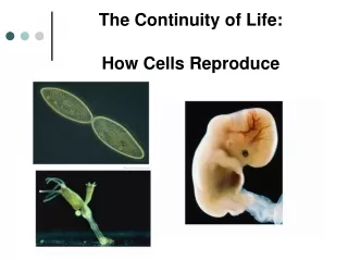
How Cells Reproduce. Chapter 9. Impacts, Issues Henrietta’s Immortal Cells. Henrietta Lacks died of cancer at age 31, but her cells ( HeLa cells) are still growing in laboratories. 9.1 Overview of Cell Division Mechanisms.

E N D
How Cells Reproduce Chapter 9
Impacts, IssuesHenrietta’s Immortal Cells • Henrietta Lacks died of cancer at age 31, but her cells (HeLa cells) are still growing in laboratories
9.1 Overview of Cell Division Mechanisms • Individual cells or organisms produce offspring by the process of reproduction • When a cell reproduces, each descendent receives information coded in DNA, and enough cytoplasm to begin operating
Mitosis, Meiosis, and the Prokaryotes • Eukaryotic cells • Mitosis copies DNA and divides a nucleus, producing two identical nuclei • Meiosis is a nuclear division that produces haploid gametes for sexual reproduction • Prokaryotic cells reproduce asexually by prokaryotic fission
Key Points About Chromosome Structure • Each species has a characteristic number of chromosomes that differ in length and shape • Each consists of one double strand of DNA • After duplication, each consists of two double strands (sister chromatids) that remain attached to each other at a centromere until late in nuclear division
one chromosome (unduplicated) one chromatid its sister chromatid one chromosome (duplicated) Fig. 9-2, p. 142
Key Points About Chromosome Structure • A chromosome consists of DNA that is wrapped around proteins (histones) and condensed • Each histone and the DNA wrapped around it make up a nucleosome, the smallest unit of structural organization in chromosomes
centromere A Duplicated human chromosome in its most condensed form. If this chromosome were actually the size shown in the micrograph, its two DNA strands would stretch out about 800 meters (0.5 miles). Fig. 9-3a, p. 143
B When a chromosome is at its most condensed, the DNA is packed into tightly coiled coils. multiple levels of coiling of DNA and proteins C When the coiled coils unwind, a molecule of chromosomal DNA and its associated proteins are organized as a cylindrical fiber. fiber D A loosened fiber shows a “beads-on-a-string” organization. The “string” is the DNA molecule; each “bead” is one nucleosome. beads on a string DNA double helix core of histones E A nucleosome consists of part of a DNA molecule looped twice around a core of histone proteins. nucleosome Fig. 9-3 (b-e), p. 143
B When a chromosome is at its most condensed, the DNA is packed into tightly coiled coils. centromere multiple levels of coiling of DNA and proteins C When the coiled coils unwind, a molecule of chromosomal DNA and its associated proteins are organized as a cylindrical fiber. A Duplicated human chromosome in its most condensed form. If this chromosome were actually the size shown in the micrograph, its two DNA strands would stretch out about 800 meters (0.5 miles). fiber D A loosened fiber shows a “beads-on-a-string” organization. The “string” is the DNA molecule; each “bead” is one nucleosome. beads on a string DNA double helix core of histones E A nucleosome consists of part of a DNA molecule looped twice around a core of histone proteins. nucleosome Stepped Art Fig. 9-3 (b-e), p. 143
G1 S Interval of cell growth before DNA replication (chromosomes unduplicated) Interval of cell growth when the DNA is replicated (all chromosomes duplicated) G2 Interval after DNA replication; the cell prepares to divide cytoplasmic division; each descendant cell enters interphase Telophase G2 Anaphase Metaphase Prophase Interphase ends for parent cell Stepped Art Fig. 9-4, p. 144
9.1 Key Concepts:Chromosomes and Dividing Cells • Individuals have a characteristic number of chromosomes in each of their cells • The chromosomes differ in length and shape, and they carry different portions of the cell’s hereditary information • Division mechanisms parcel out the information into descendent cells
9.2 Introducing the Cell Cycle • Cell cycle • A sequence of three stages (interphase, mitosis, and cytoplasmic division) through which a cell passes between one cell division and the next
Interphase • Interphase consists of three stages, during which a cell increases in size, doubles the number of cytoplasmic components, and duplicates its DNA • G1: Interval of cell growth and activity • S: Interval of DNA replication (synthesis) • G2: Interval when the cell prepares for division
Interphase and the Life of a Cell • Most cell activities take place during G1 • Control mechanisms work at certain points in the cell cycle; some can keep cells in G1 • Loss of control may cause cell death or cancer
Mitosis and the Chromosome Number • Mitosis produces two diploid nuclei with the same number and kind of chromosomes as the parent • Chromosome number • The sum of all chromosomes in a type of cell • Human cells have 46 chromosomes paired in 23 sets (diploid number) • Pairs have the same shape and information about the same traits (except sex chromosomes XY)
Mitosis and the Chromosome Number • Bipolar spindle • A dynamic network of microtubules that forms during nuclear division • Grows into the cytoplasm from opposite poles of the cell and attaches to duplicated chromosomes • Microtubules from opposite poles attach to different sister chromatids and separate them
After mitosis and cytoplasmic division, the two new cells each have one (unduplicated) chromosome. Both new cells start life in G1 of interphase. mitosis, cytoplasmic division An undupli- cated chromosome in a cell in G1 of interphase. The same chromosome, duplicated in S. The cell is now in G2 of interphase. Fig. 9-5b, p. 145
9.2 Key Concepts:Where Mitosis Fits in the Cell Cycle • A cell cycle starts when a new cell forms by division of a parent cell, and ends when the cell completes its own division • A typical cell proceeds through intervals of interphase, mitosis, and cytoplasmic division
9.3 A Closer Look at Mitosis • When a nucleus divides by mitosis, each new nucleus has the same chromosome number as the parent cell • There are four main stages of mitosis: prophase, metaphase, anaphase, and telophase
Prophase • Prophase • Chromosomes condense • Microtubules form a bipolar spindle • Nuclear envelope breaks up • Microtubules attach to the chromosomes • Centrosome • A region near the nucleus that organizes spindle microtubules; usually includes two centrioles
Metaphase and Anaphase • Metaphase • All duplicated chromosomes line up midway between the spindle poles • Anaphase • Microtubules separate the sister chromatids of each chromosome and pull them to opposite spindle poles
Telophase • Telophase • Two clusters of chromosomes reach the spindle poles • A new nuclear envelope forms around each cluster • Two new nuclei are formed, each with the same chromosome number as the parent cell
A Early Prophase Mitosis begins. In the nucleus, the chromatin begins to appear grainy as it organizes and condenses. The centrosome is duplicated. Fig. 9-6 (2a), p. 147
B Prophase The chromosomes become visible as discrete structures as they condense further. Microtubules assemble and move one of the two centrosomes to the opposite side of the nucleus, and the nuclear envelope breaks up. Fig. 9-6 (2b), p. 147
C Transition to Metaphase The nuclear envelope is gone, and the chromosomes are at their most condensed. Microtubules of the bipolar spindle assemble and attach sister chromatids to opposite spindle poles. Fig. 9-6 (2c), p. 147
D Metaphase All of the chromosomes are aligned midway between the spindle poles. Microtubules attach each chromatid to one of the spindle poles, and its sister to the opposite pole. Fig. 9-6 (2d), p. 147
E Anaphase Motor proteins moving along spindle microtubules drag the chromatids toward the spindle poles, and the sister chromatids separate. Each sister chromatid is now a separate chromosome. Fig. 9-6 (2e), p. 147
F Telophase The chromosomes reach the spindle poles and decondense. A nuclear envelope begins to form around each cluster; new plasma membrane may assemble between them. Mitosis is over. Fig. 9-6 (2f), p. 147