Download

1 / 21
361 likes | 1.39k Views
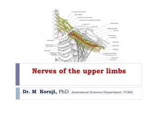
Nerves of the thorax. Phrenic nerves. - 0rigin:- ventral rami of C4 mainly and C3,C5. - It is a mixed nerve (has motor, sensory fibers). Motor:- supply diaphragm. Sensory:- supply pericardium (fibrous and parietal layer of serous),

E N D
Phrenic nerves - 0rigin:- ventral rami of C4 mainly and C3,C5. - It is a mixed nerve (has motor, sensory fibers). Motor:- supply diaphragm. Sensory:- supply pericardium (fibrous and parietal layer of serous), - Parietal pleura (mediastinal and central part of diaphragmatic). - Branches to mediastinum
Course of right phrenic Nerve (venous) - It descends on the right side of: a. Right innominate. b. Superior vena cava. c. Right atrium (separated form it by the pericardium). d. Inferior vena cava. In front of the root of the right lung. - Then, it passes through the IVC opening of the diaphragm to be distributed on the inferior surface of the diaphragm.
Course of left phrenic Nerve (arterial) - It ↓ between the left common carotid and left subclavian arteries. - ↓in front of the arch of aorta and crosses also the left vagus. - ↓ on the left side of the left ventricle (separated form it by the pericardium). As it descends it lies in front of the root of the left lung. Then, it pierces the left copula of the diaphragm to be distributed on the inferior surface of the diaphragm.
Vagi nerves • Vagus is the tenth (X) cranial nerve. - Originates from medulla oblongata, - Descends all the way down from the brainstem to the colon .
Right vagus nerve Course: - ↓ Anterior to right subclavian artery - ↓ on Rt side of trachea. - ↓ Behind Rt main bronchus. - Breaks to form posterior pulmonary plexus. • Reforms then breaks to form esophageal plexus. • Reforms to make posterior gastric nerve. • Enters abdomen through esophageal opening of diaphragm.
Left vagus nerve Course: - ↓ between LCC , LSC. - Crosses left surface of aortic arch. - ↓ behind left main bronchus. - Breaksto form posterior pulmonary plexus. • Reforms then breaks to form esophageal plexus. • Reforms to make anterior gastric nerve. • Entersabdomen by passing through esophageal opening of diaphragm.
Branches: 1. Left recurrent laryngeal nerve: It recurs below the arch of the aorta to ascend in the groove between theleft side of the oesophagus and trachea. 2. Filaments to four plexuses They arise from both sides to the following plexuses: a. Cardiac plexus. b. Anterior pulmonary plexus c. Posterior pulmonary plexus. d. Oesophageal plexus.
Sympathetic trunk (Chain) The sympathetic trunk is formed of the sympathetic ganglia and the communications between them. It is present in the thorax and extends up to the neck and down to the abdomen and pelvis. The thoracic part contains 12 ganglia (someimes 11 due to fusion of ganglia 11 and 12). The first thoracic ganglion may be fused with the inferior cervical one to (75% - 80% of people) form the Stellate ganglion. Sites: the ganglia are present posteriorly in the thorax, close to the heads of the ribs or at the sides of vertebral bodies.
Branches of the sympathetic trunk 1. Communicating branches to the corresponding spinal nerves (white rami communicants = preganglionic fibres and grey rami communicants = postganglionic fibres). 2. Oesophageal branches (1st - 4th ganglia). 3. Pulmonary branches (1st - 4th ganglia). 4. Cardiac branches (1st - 4th ganglia). 5. Splanchnic branches: a.Greater splanchnic nerve (5th - 9th ganglia). b. Lesser splanchnic nerve (10th - 11th ganglia). c. Lowest splanchnic nerve (12th ganglia).
Innervation of the viscera of the thorax(nerve plexuses of the thorax) 1- Cardiac Plexuses: - They are autonomic plexuses (sympathetic and vagi) which supply the heart. A- Superficial Cardiac Plexus: Site: Below the arch of the aorta. On the right side of ligamentum arteriosum. Formation: Cardiac branch of the left superior cervical sympathetic ganglia. - Cardiac branches of the left vagus. Distribution: 1. Deep part of the cardiac plexus. 2. Right coronary plexus. 3. Left anterior pulmonary plexus.
B- Deep Cardiac Plexus:Site: In front of the bifurcation of the trachea.Formation: - Cardiac branches of all cervical sympathetic ganglia (except left superior).- Cardiac branches of the upper (1- 4) thoracic sympathetic ganglia.- Cardiac branches of the Right vagus.- Cardiac branches of right and left recurrent laryngeal nerves.Distribution:1. Superficial cardiac plexus.2. right and left anterior pulmonary plexuses.3. Right and left coronary plexuses.4. Right and left atria.
2- Coronary Plexuses:- Site: They surround the coronary arteries and their branches.- Formation: They receive branches from the cardiac plexuses. 1. Right coronary plexus (along the right coronary artery)2. Left coronary plexus (along the left coronary artery)- Function:- Sympathetic fibers increase heart rate and cause vasodilatation of the coronaries.- Parasympathetic fibers decrease heart rate and cause vasoconstriction of the coronaries.
3- Pulmonary Plexuses: Site: There are anterior and posterior to the hilum of each lung ( in relation to the main bronchi). Formation: They are extensions from the cardiac plexus. A. Anterior pulmonary plexus(smaller) formed by: - Vagi nerves. - Superficial cardiac plexus.
B. Posterior pulmonary plexus (larger) - Formed by: 1- Vagi nerves. 2- Deep cardiac plexus. 3- Thoracic sympathetic ganglia (2 - 5). 4- Left recurrent laryngeal nerve. - Function: - Sympathetic fibers cause broncho-dilatation and decreased secretions. - Parasympathetic fibers cause broncho-constriction and increased secretions.
4- Oesophageal Plexus: Site: It lies around the wall of the oesophagus in the posterior mediastinum. Formation: - It is formed by both vagi and filaments of the sympathetic trunk (upper 5 thoracic ganglia, and the greater splanchnic nerve). Function: - Sympathetic fibers inhibit peristalsis (muscle relaxation) and decrease secretions. - Parasympathetic fibers stimulate peristalsis and increases secretions.