Download
1 / 22
310 likes | 1.23k Views
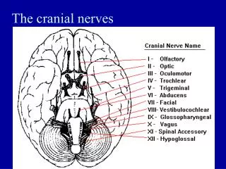
EXAMINATION OF THE CRANIAL NERVES. OLFACTORY NERVE (I). Test with alcowipes, coffee etc. Unilateral anosmia may be significant Bilateral anosmia: commonest cause viral Classical pathology:olfactory groove meningioma Basal skull fractures another potential cause (unilateral or bilateral).

E N D
OLFACTORY NERVE (I) • Test with alcowipes, coffee etc. • Unilateral anosmia may be significant • Bilateral anosmia: commonest cause viral • Classical pathology:olfactory groove meningioma • Basal skull fractures another potential cause (unilateral or bilateral)
OPTIC NERVE (II) • Visual acuity • Visual fields to confrontation • Pupillary reflexes (II and III) • Fundoscopy (papilloedema, optic atrophy, retinitis pigmentosa)
VISUAL ACUITY • CORRECTED (ie brain not lens) • Each eye separately • Snellen charts for distance and near vision reading charts for near vision • Best approximation: small print (or equivalent) at normal reading distance • If unable, finger counting, hand movements, perception of light
VISUAL FIELDS • Often forgotten but very important • First do a bilateral screening test: will uncover the majority of significant visual field defects immediately • Go on to check each eye separately, ask about scotomata • Mention checking for blind spot enlargement
COMMON FIELD DEFECTS • HOMONOMOUS HEMIANOPIA: lesion posterior to the optic chiasm (eg posterior cerebral artery territory infarction) • BITEMPORAL HEMIANOPIA: lesion at the optic chiasm (eg pituitary tumour) • BLINDNESS ONE EYE: lesion in eye, retina or optic nerve
PUPILLARY RESPONSES • Light reflex is the clinically significant one • Afferent limb = II, efferent limb = III • Look at pupillary sizes • Direct and consensual response • Look for afferent pupillary defect (optic nerve lesion)
PUPILLARY ABNORMALITIES • One large pupil: IIIrd nerve palsy, iris problem (eg traumatic midriasis), unilateral dilator eye drops • Small pupil: Horner’s syndrome, Argyll-Roberston pupil (small, irregular, reacts to accommodation but not to light) • Bilateral small pupils: drugs (opiates), pontine lesion (haemorrhage)
HORNER’S SYNDROME • Oculosympathetic paralysis • A good lateralising sign but a poor localising sign • Ptosis, miosis and sometimes unilateral anhydrosis of face • Look especially at neck, supraclavicular fossa and hand (Pancoast’s tumour)
Eye movements (III, IV and VI) • IV: TROCHLEAR NERVE (supplies superior oblique muscle) • VI: ABDUCENT NERVE (supplies lateral rectus muscle) • III: OCULOMOTOR NERVE: all other extraocular muscles, also carries parasympathetic (constrictor) fibres to pupil, and fibres to levator palpebrae superioris
EYE MOVEMENTS • Look at eyes in primary position of gaze • IIIrd nerve palsy: eye often ‘down and out’ • VI nerve palsy: often eyes convergent (unopposed medial rectus) • Look at pupils • Look for ptosis
EYE MOVEMENTS • Follow a moving object (finger, end of tendon hammer) and ask for any symptomatic diplopia • Determine position/s causing maximum diplopia • Ask about separation of images (horizontal or oblique) • Check diplopia is BINOCULAR
TYPICAL EXAM CASES • IIIrd nerve palsy: ptosis, eye ‘down and out’, diplopia in all except one direction of gaze, may have dilated pupil ( a ‘surgical’ IIIrd nerve palsy • VI nerve palsy: eye convergent, diplopia on lateral gaze only, horizontally separated images
CAUSES OF COMPLEX OPTHALMOPLEGIA • Dysthyroid eye disease • Myasthenia gravis (look for fatiguability of diplopia and ptosis) • Mitochondrial disorders
INTERNUCLEAR OPHTHALMOPLEGIA • Nystagmus in the abducting eye • Failure of adduction of the other eye • Both eyes move normally when tested individually • Lesion in the MEDIAL LONGITUDINAL FASICULUS (on the side WITHOUT nystagmus • Can be bilateral
TRIGEMINAL NERVE (V) • Most important function is sensory • Ophthalmic, maxillary and mandibular divisions • Test with light touch and pinprick in all 3 divisions, comparing each side • Corneal reflexes (afferent limb V, efferent limb VII) • Know something about trigeminal neuralgia (examination is normal in these cases)
FACIAL NERVE (VII) • Supplies the muscles of the face • DIFFERENTIATE AN UPPER MOTOR NEURON FROM A LOWER MOTOR NEURON LESION • Upper motor neuron lesion: milder, spares the forehead, no Bell’s phenomenon
VESTIBULOCOCHLEAR NERVE (VIII) • For clinical examination purposes, forget the vestibular element • Check hearing approximately in each ear • If reduced, determine whether conductive (BC >AC) or sensorineural (AC>BC) deafness
GLOSSOPHARYNGEAL (IX) AND VAGUS (X) • Tested together • Speech, palate, cough, swallow, (gag reflex) • Bulbar palsy: bilateral LMN lesions of IX and X: poor palatal movement, nasal speech, nasal regurgitation of fluids • Pseudobulbar palsy: bilateral UMN lesions: ‘hot potato’ speech, no nasal regurgitation, additional frontal lobe signs
ACCESSORY NERVE (XI) • Cranial and spinal roots • Cranial roots: sternocleidomastoid (note direction of head turn) • Spinal roots: trapezius (shoulder shrug)
HYPOGLOSSAL NERVE • Movement of the tongue • Look for wasting and fasiculation of the tongue • Deviation of tongue on protrusion • Tongue movements including power