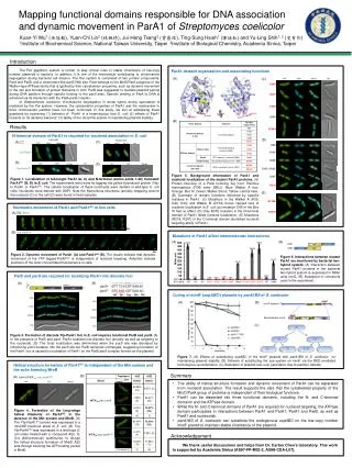
Exploring Functional Domains of ParA1Scoe in Streptomyces coelicolor and Its Cytoskeletal Properties
10 likes | 130 Views
This study investigates the ParA1Scoe protein from Streptomyces coelicolor, focusing on its domain organization, nucleoid localization, and dynamic movement in E. coli. Through mutational analyses, we show that specific mutations in the Walker A and B boxes affect nucleoid targeting, while the C-terminal domain remains unaffected. Our findings confirm that ParA1Scoe shares similarities with other ParA proteins and highlight the independent roles of its domains in nucleoid association and cytoskeletal dynamics.

Exploring Functional Domains of ParA1Scoe in Streptomyces coelicolor and Its Cytoskeletal Properties
E N D
Presentation Transcript
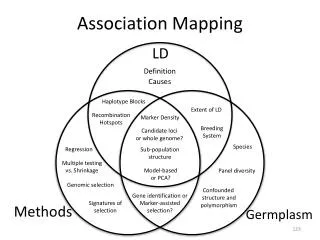
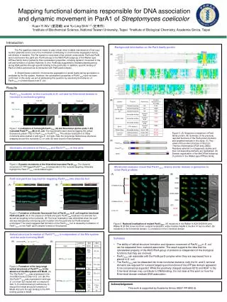
(C) N-terminus D152 K39 K44 G40 180º R247 R247 R218 R218 Walker A 44 40 39 BoxIV Walker B 68 152 218 247 2 4 6 8 10 Mutational analyses reveal that ParA1Scoeshares similar domain organization to other ParA proteins 12 14 16 18 20 22 24 (A) (B) YFP DAPI merge DIC YFP DAPI merge DIC R8E K39E R19E G40V In Walker A box R26E Plac Plac parS parS* (parB silent mutation) yfp parA1Scoe parB yfp parA1Scoe parB K44E R19E+R26E Q29E In Walker B box D154A (C) R218E In box IV D68A R247E Figure 6. Nucleoid localization of mutant ParA1Scoe. (A) mutations in the Walker A (K39/G40/K44) and Walker B (D154) boxes lost their nucleoid localizaiton, while mutation Asp68 in the Box VI has no effect. (B) mutations in the N-terminal domain. (C) mutations in the C-terminal domain time image Cell count (n) Acknowledgement This work is supported by Academia Sinica (AS97-FP-M02-2). Cell count (n) Mapping functional domains responsible for DNA association and dynamic movement in ParA1 of Streptomyces coelicolor Kuan-Yi Wu1 (巫冠毅) and Yu-Ling Shih1, 2 (史有伶) 1Institute of Biochemical Science, National Taiwan University, Taipei2Institute of Biological Chemistry, Academia Sinica, Taipei Introduction Background information on the ParA family protein The Par (partition) system is known to play critical roles in stable maintenance of low-copy number plasmids and is one of the mechanism contributing to chromosome segregation during cell division in bacteria. The Par system is comprised of two protein components, ParA and ParB, and a centromere-like parS site. ParA belongs to the MinD/ParA subgroup of the Walker-type ATPase family that is typified by their cytoskeleton properties, including dynamic movement in the cell and formation of protein filaments in vitro. ParB was suggested to mediate plasmid pairing during DNA partition through specific binding to the parS sites. In addition, specific binding of ParA to DNA is achieved via its interaction with ParB-parS complex. In Streptomyces coelicolor, chromosome segregation in aerial hypha during sporulation is mediated by the Par system. However, the cytoskeleton properties of ParA1Scoehave not been confirmed. In this study, we aim at addressing this question by examining the behavior of ParA1Scoe in a heterologous host E. coli. . (A) Results ParA1Scoe localizes to the nucleoids in E. coli and its N-terminal domain is involved in nucleoid-targeting (B) Live cell Fixed cell (A) (C) YFP DAPI merge DIC YFP DAPI merge DIC Yfp::ParA1Scoe YFP DIC merge (B) (D) Yfp::ParA1NCScoe Figure 1. Localization of full-length ParA1Scoe (A) and N-terminal (amino acids 1-32) truncated ParA1NCScoe (B) in E. coli. The experiments were done by tagging the yellow fluorescent protein (Yfp) to ParA1Scoe or ParA1NCScoe. The cellular localization of these constructs were studied in wild-type E. coli cells. Note the long-range filamentous structures wrapping around the nucleoids (C) or cells (D) were found in fixed samples. Figure 5. (A) Sequence comparison of ParA family protein. (B) Summary of the previosly reported functions of the ParA protein domains and targeted residues in this study. (C) Two views of the protein structure of Soj from Thermus thermophilus (PDE entry 2BEJ). Residues selected in mutagenesis studies and their corresponding domains are highlighted. (D) Domain organization of the MinD/ParA subgroup of proteins in the Walker-type ATPase family. (D) ParA1Scoe Stochastic movement of ParA1Scoeand ParA1NCScoe in live cells ParA1NCScoe MinD 0s Figure 2. Dynamic movement of the N-terminal truncated ParA1Scoe. The dynamic movement of YFP taggedParA1NCScoeis independent of the nucleoid targeting. Asterisks highlight the ParA1NCScoe concentrated region. ParB and parS are required for targeting ParA1Scoeinto discrete foci (A) (B) YFP YFP DAPI DAPI merge merge DIC DIC Figure 3. Formation of discrete fluorescent foci ofParA1Scoe in E. coli requires functional ParB and parS.(A) In the presence of ParB and parS, ParA1Scoelocalized in to discrete foci as well as targeting to the nucleoids. (B) The focal localization was diminished when the parS site was disrupted by introducing point mutation into the parS site but ParB remained unchanged. The result suggests that formation of the ParA1Scoe foci is caused by nucleation of ParA1Scoe on the ParB- parS complex formed on the plasmid. Helical structure formation ofParA1NCScoe is independent of the Min system and the actin homolog MreB Summary • The ability of helical structure formation and dynamic movement of ParA1Scoein E. coli can be separated from nucleoid association. The result supports the idea that the cytoskeletal property of the MinD/ParA group of proteins is independent of the biological functions that they are involved. • ParA1Scoe can associate with the ParB-parS complex when they are expressed from a plamid in E. coli. • The ParA1Scoe can be dissected into three functional domains: both the N- and C-terminal domains are required for nucleoid targeting and functions of the ATPase domain appeared to be conserved as expected. While the positively charged residues R218 and R247 in the C-terminal domain may contribute to DNA binding, it is not clear at this point on how the N-terminal domain mediate DNA association. (A) (B) ΔminCDE/Plac- yfp::parA1NCScoe YFP DIC merge Figure 4. Formation of thelong-range helical structures of ParA1NCScoe in the absence of the Min system and MreB. (A) The Yfp-ParA1NCScoe was expressed in the minCDE knockout strain of E. coli. (B)The Yfp-ParA1NCScoe was expressed in wild-type E. coli strain but treated with a compound A22, S-(3,4 dichlorobenzyl) isothiourea, to disrupt the helical structure formation of MreB. A22 acts through binding to the ATP-binding pocket in MreB.