Forefoot Fractures
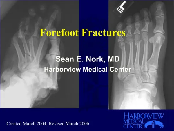
Forefoot Fractures. Sean E. Nork, MD Harborview Medical Center. Created March 2004; Revised March 2006. Foot Trauma and Outcomes. Turchin et al, JOT, 1999 28 patients: Polytrauma +/- foot injury Age, gender, ISS matched Results SF-36 5/8 components worse with foot injury

Forefoot Fractures
E N D
Presentation Transcript
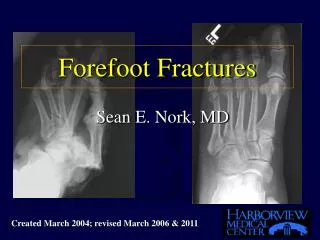
Forefoot Fractures • Sean E. Nork, MD • Harborview Medical Center Created March 2004; Revised March 2006
Foot Trauma and Outcomes • Turchin et al, JOT, 1999 • 28 patients: Polytrauma +/- foot injury • Age, gender, ISS matched • Results SF-36 5/8 components worse with foot injury • WOMAC All 3 components worse with foot injury Jurkovich et al, JT, 1995 Highest Sickness Impact Profile (SIP) @ 6 & 12 months Patients with foot trauma (compared to other lower extremity injuries)
Foot Function • Hindfoot: Shock absorption, propulsion, deceleration • Midfoot: Controls relationship between hindfoot and forefoot • Forefoot: Platform for standing and lever for push off
Forefoot Function • Platform for weight bearing • Lever for propulsion
Anatomy • First Metatarsal • Shorter & wider • Bears 1/3 body weight • Tendon attachments: (Tibialis Anterior & Peroneus Longus) • Tibialis Anterior: varus, supination, elevation • Peroneus Longus: valgus, pronation, depression Lesser Metatarsals More mobile medial to lateral Bear 1/6 weight each Intermetatarsal ligaments (2-3, 3-4, 4-5)
Sesamoids • Medial (tibial) & Lateral (fibular) • Within FHB tendons • Articulate with 1st MT head • Weight bearing through sesamoids Tibial Sesamoid:Tibial FHB Abductor Hallucis Fibular Sesamoid:Fibular FHB Adductor Hallucis Deep Tverse MT ligament
Phalanges • Great toe (2) • Lesser toes (3) • FDB attaches @ intermediate • FDL/EDL attaches @ distal
Biomechanics • Metatarsal heads in contact with floor 60-80% of stance phase • Toes in contact with floor 75% • of stance phase Cavanagh, PR, F&A, 1987 Hughes, J, JBJS[Br], 1990
Cross-sectional Geometry of the Human Forefoot • Griffin & Richmond, Bone, 2005 • Examines the relationship between external loads during walking & running and the geometrical properties of the human forefoot • Metatarsals 2-4 are the weakest in most cross-sectional geometric properties • Metatarsal 2 (and 3 to a lesser extent) experience high peak pressures; this may explain the preponderance of stress fractures in these metatarsals
Mechanism • Industrial accidents • MVA (airbags) • Indirect (twisting injuries) • Other
Physical Examination • Gross deformity • Dislocations • Sensation • Capillary refill • Foot Compartments
Radiographs • Foot trauma series • AP/lat/oblique • Don’t forget oblique • Sesamoid view • Tangential view (MT heads) • Contralateral foot films (comparison) • CT Scan (occasionally)
Treatment Principles • Hindfoot: Protect subtalar, ankle and talonavicular joints • Midfoot: restore length and alignment of medial and lateral “columns” • Forefoot: Even weight distribution
Treatment • Border Rays • First metatarsal • Fifth metatarsal • Dislocations • Multiple metatarsal shafts • Intraarticular fractures
First MT Shaft Fractures • Nondisplaced • Consider conservative treatment • Immobilization with toe plate • Displaced • Most require ORIF • Strong muscle forces (TA, PL) • Deformity common • Bears 2/6 body weight ORIF Plate and screws Anatomically reduce May cross first MTP joint (temp)
First MT Base Fractures • Articular injuries • Frequently require ORIF • Fixation: • Spans TMT • Doesn’t span TMT • Temporarily Spans TMT
36 year old males/p MVCActive Note articular comminution
After ORIF Fixation Strategy Direct ORIF of comminuted first MT base fractre Temporary spanning across first TMT joint
43 year old male injured in a MVCObserve the articular segment impaction of the base of the first.The first MT is shortened and dorsally displaced while the plantar ligaments remain attached.
The patient underwent ORIF of the base of the first metatarsal with spanning of the first TMT, given the level of comminution observed. Additionally, temporary spanning external fixation was used.
Radiographic appearance at 3 months after removal of the external fixator and metatarsal neck k-wire fixations.
Non-displaced Metatarsal Fractures 2-4 • Single metatarsal fractures • Treatment usually nonoperative • Symptomatic: hard shoe vs AFO vs cast vs elastic bandage • Multiple metatarsal fractures • Usually symptomatic treatment (as above) • May require ORIF if other associated injuries
Minimally Displaced Lesser Metatarsal Fractures • Zenios et al, Injury 2005 • Prospective and randomized (n=50) • Case vs elastic support bandage • MINIMALLY DISPLACED fractures • Higher AOFAS mid-foot scores at 3 months and less pain if treated with an elastic support bandage.
Displaced Metatarsal Shaft Fractures • Sagittal plane displacement & angulation • is most important. • Reestablish length, rotation, & declination • Dorsal deformity can produce transfer metatarsalgia • Plantar deformity can produce increased load at affected metatarsal Treatment Options Closed Reduction Intramedullary pinning with k-wire (0.054” or 0.062”) Pinning of distal segment to adjacent metatarsal ORIF with dorsal plate fixation
This patient sustained an open second metatarsal fracture in a crush injury. Given the soft tissue injury and continued pressure on the dorsal skin, operative fixation was elected.
Fixation consisted of a dorsal 2.0 mm plate application after appropriate irrigation of the open fracture.
This patient was treated with ORIF of multiple metatarsal fractures (3,4,5) through a dorsal approach. Fixation consisted of a 2.7 mm DCP on the fifth and 2.0 mm plates on the third and fourth metatarsals.
Medullary K-wires in Lesser MTs • Exit wire distally through the proximal phalanx • Plantar wire exit may produce a hyperextension deformity of the MTP ST Hansen, Skeletal Trauma
This patient sustained multiple metatarsal neck fractures (2, 3, 4) and a dislocation of the fifth MTP joint. Note the lateral translation, lateral angulation, and the displacement on the lateral radiograph. Compliments: Daphne Beingessner, MD
Stabilization consisted of closed reduction and percutaneous pin fixation of the multiple metatarsal fractures and closed reduction of the fifth MTP dislocation. Note the location and trajectory of the K-wires. Compliments: Daphne Beingessner, MD
Following healing and removal of the pins, good alignment of the forefoot is demonstrated on the multiple radiographic views. Compliments: Daphne Beingessner, MD
Stress Fractures of Metatarsals 2 - 4 • Identify Cause • First ray hypermobility • Short first ray • Tight gastrocnemius • Long metatarsal • Treatment • Treat cause if identifiable • If overuse, activity restriction • Reserve ORIF for displaced fractures
Metatarsal Neck Fractures • Usually displace plantarly • May require reduction and fixation: • Closed reduction and pinning • Open reduction and pinning • ORIF (dorsal plate)
This patient sustained multiple metatarsal neck fractures after an MVA. Note additional fractures at the first and fifth metatarsals
Medullary wire fixation of metatarsal neck fractures 2, 3, 4 Compliments of S.K. Benirschke
Metatarsal Head Fractures • Unusual • Articular injuries • May require ORIF • (especially if first MT) Circular saw injury to the articular surface of the first MT head
Fifth Metatarsal Fractures • Mid diaphyseal fractures • Stress fractures (proximal diaphysis) • Jones fractures (metadiaphyseal jxn) • Tuberosity fractures
Proximal Fifth Metatarsal FracturesDameron, TB, JAAOS, 1995 • Zone 1 cancellous tuberosity • insertion of PB & plantar fascia • involve metatarsocuboid joint • Zone 2 distal to tuberosity • extend to 4/5 articulation • Zone 3 distal to proximal ligaments • usually stress fractures • extend to diaphysis for 1.5 cm
Proximal Fifth Metatarsal FracturesDameron, TB, JAAOS, 1995 • Relative Frequency • Zone 1 93% • Zone 2 4% • Zone 3 3%
Fifth Metatarsal Blood Supply Shereff, M et al, F&A, 1991 Fresh leg specimens (after BKA) (n = 15) Extraosseus circulation: dorsal metatarsal artery plantar metatarsal artery fibular plantar marginal artery Intraosseus circulation: Nutrient artery Metaphyseal vessels Periosteal complex • Smith, J et al, F&A, 1992 • Cadaver Arterial Injection Study (n = 10) • Nutrient artery with intramedullary branches (retrograde flow to proximal fifth metatarsal) • Multiple metaphyseal arteries • Conclusions: Fracture distal to the tuberosity disrupts the nutrient arterial supply and creates relative avascularity
Fifth Metatarsal Blood Supply Smith et al, Foot Ankle 1993
Zone 1 Fractures: Tuberosity • Etiology • Avulsion from lateral plantar aponeurosis • (Richli & Rosenthal, AJR, 1984) • Treatment • Symptomatic • Hard shoe • Healing usually uneventful • (Dameron, T, JBJS, 1975) Lawrence, SL, Foot Ankle, 1993
Zone 1 Fractures: TuberosityWeiner, et al, F & A Int, 1997 • 60 patients • Randomized to short leg cast vs soft dressing only • Weight bearing in hard shoe in all • Healing in 44(average) - 65(all) days • Soft dressing only: shorter recuperation (33 vs 46 days) and similar foot score (92 vs 86) • Conclusions: Faster return to function without compromising radiographic union or clinical outcome in patients treated without casting.
Zone 2 Fractures: Metadiaphyseal • Treatment Controversial • Union frequently a concern • Early weight bearing associated with increased nonunion(Torg, Ortho, 1990; Zogby, AJSM, 1987) Nondisplaced Fractures: Treatment Cast with non weight bearing (Shereff, Ortho, 1990; Heckman, 1984; Hens, 1990; Lawrence, 1993) Cast with weight bearing (Kavanaugh, 1978; Dameron, 1975)
Zone 2 Fractures: Metadiaphyseal • Operative Treatment • Medullary Screw Stabilization • (Delee, 1983; Kavanaugh, 1978; Dameron, 1975) • Bone Graft Stabilization • (Dameron, 1975; Hens, 1990; Torg, 1984)
Zone 2 Fractures: Metadiaphyseal • Operative Treatment • Medullary Screw Stabilization • Bone Graft Stabilization Lehman, Foot Ankle 1987
MTP Joint Injuries • Sprains • “Turf Toe”: hyperextension with injury to thee plantar plate • Hyperflexion sprains • Dislocations